Development of the nervous system: Difference between revisions
imported>OAbot m Open access bot: doi updated in citation with #oabot. |
imported>JCW-CleanerBot m clean up, removed: (New York, N.Y.) |
||
| (One intermediate revision by one other user not shown) | |||
| Line 1: | Line 1: | ||
{{Short description|Processes which grow and shape an organism's nervous tissue over its lifetime(s)}} | {{Short description|Processes which grow and shape an organism's nervous tissue over its lifetime(s)}} | ||
{{About|neural development in all types of animals, including humans|information specific to the human nervous system|Development of the nervous system in humans}} | |||
{{cs1 config|name-list-style=vanc|display-authors=6}} | {{cs1 config|name-list-style=vanc|display-authors=6}} | ||
{{Organ system development}} | {{Organ system development}} | ||
The ''' | The construction of the nervous system is one of the most complex processes in embryology. '''Development of the nervous system''', or '''neural development''' ('''neurodevelopment'''), refers to the processes that generate, shape, and reshape the [[nervous system]] of animals, from the earliest stages of [[embryonic development]] to adulthood. In vertebrates, it begins with the formation of the neural tube from the ectoderm via neurulation. This tube then differentiates into the brain and spinal cord through regionalization and patterning by morphogen gradients. Subsequent stages include neurogenesis (the birth of neurons) neuronal migration, axon guidance, synaptogenesis, and extensive activity-dependent refinement to produce functional neural circuits'''.''' This field of neural development draws on both [[neuroscience]] and [[developmental biology]] to describe and provide insight into the cellular and molecular mechanisms by which complex nervous systems develop, from [[nematode]]s and [[Drosophila melanogaster|fruit flies]] to [[mammals]]. | ||
Defects in neural development can lead to malformations such as [[holoprosencephaly]], and a wide variety of [[neurological disorder]]s including [[paresis|limb paresis]] and [[paralysis]], balance and vision disorders, and [[seizure]]s,<ref name="Vet">{{cite web |title=Neurological Signs & Diseases |url=http://vetneuro.com/NeurologicalSignsDiseases/tabid/4171/Default.aspx |access-date=1 May 2020 |date=2 November 2016|archive-url=https://web.archive.org/web/20161102033420/http://vetneuro.com/NeurologicalSignsDiseases/tabid/4171/Default.aspx |archive-date=2016-11-02 }}</ref> and in [[human]]s other disorders such as [[Rett syndrome]], [[Down syndrome]] and [[intellectual disability]].<ref>{{cite web|title=Neural Tube Defects|url=https:// | Recent advances in genomics and imaging technologies, such as single-cell sequencing and live-cell microscopy, have refined our understanding of neural development at molecular and cellular levels'''.''' Techniques such as single-cell RNA sequencing allow researchers to profile gene expression in individual neural progenitors and neurons, revealing previously unknown cellular diversity during development. Defects in neural development can lead to malformations such as [[holoprosencephaly]], and a wide variety of [[neurological disorder]]s including [[paresis|limb paresis]] and [[paralysis]], balance and vision disorders, and [[seizure]]s,<ref name="Vet">{{cite web |title=Neurological Signs & Diseases |url=http://vetneuro.com/NeurologicalSignsDiseases/tabid/4171/Default.aspx |access-date=1 May 2020 |date=2 November 2016|archive-url=https://web.archive.org/web/20161102033420/http://vetneuro.com/NeurologicalSignsDiseases/tabid/4171/Default.aspx |archive-date=2016-11-02 }}</ref> and in [[human]]s other disorders such as [[Rett syndrome]], [[Down syndrome]] and [[intellectual disability]].<ref>{{cite web|title=Neural Tube Defects|url=https://medlineplus.gov/neuraltubedefects.html|access-date=6 December 2011}}</ref> | ||
== Vertebrate brain development == | == Vertebrate brain development == | ||
[[File:NSdiagram.svg|thumb|upright=2.2|Diagram of the vertebrate nervous system]] | [[File:NSdiagram.svg|thumb|upright=2.2|Diagram of the vertebrate nervous system]] | ||
{{Further|Development of the nervous system in humans}} | {{Further|Development of the nervous system in humans}} | ||
The [[vertebrate]] [[central nervous system]] (CNS) is derived from the [[ectoderm]]—the outermost [[germ layer]] of the embryo. A part of the dorsal ectoderm becomes specified to neural ectoderm – [[neuroectoderm]] that forms the [[neural plate]] along the dorsal side of the embryo.<ref name="Gilbert"/><ref name=Zhou>{{cite journal | vauthors = Zhou Y, Song H, Ming GL | title = Genetics of human brain development | journal = Nature Reviews. Genetics | volume = 25 | issue = 1 | pages = 26–45 | date = January 2024 | pmid = 37507490 | pmc = 10926850 | doi = 10.1038/s41576-023-00626-5 }}</ref> This is a part of the early patterning of the embryo (including the invertebrate embryo) that also establishes an anterior-posterior axis.<ref name="Wolpert1">{{cite book | vauthors = Wolpert L |title=Principles of development |url = https://archive.org/details/principlesofdeve0000unse_j4n3/page/n5/mode/2up |year=2015 |publisher=Oxford University Press |isbn=978-0-19-967814-3 |oclc = 914509705 | edition=Fifth}}</ref>{{sfn|Wolpert|2015|pages=522–526}} The neural plate is the source of the majority of neurons and glial cells of the CNS. The [[neural groove]] forms along the long axis of the neural plate, and the neural plate folds to give rise to the [[neural tube]].<ref>{{cite book| vauthors = Saladin K |title=Anatomy & Physiology The Unity of Form and Function|year=2011|publisher=McGraw Hill|location=New York|isbn= 978-0-07-337825-1|page=514}}</ref> This process is known as [[neurulation]].<ref>{{cite book | vauthors = Schoenwolf GC, Smith JL | chapter = Mechanisms of Neurulation |date=2000 | title = Developmental Biology Protocols: Volume II | series = Methods in Molecular Biology |pages=125–134 | veditors = Tuan RS, Lo CW |place=Totowa, NJ |publisher=Humana Press |language=en |doi=10.1385/1-59259-065-9:125 |isbn=978-1-59259-065-0 |volume=136 |pmid=10840705 }}</ref> When the tube is closed at both ends it is filled with embryonic cerebrospinal fluid.<ref name="Gato">{{cite journal | vauthors = Gato A, Alonso MI, Martín C, Carnicero E, Moro JA, De la Mano A, Fernández JM, Lamus F, Desmond ME | title = Embryonic cerebrospinal fluid in brain development: neural progenitor control | journal = Croatian Medical Journal | volume = 55 | issue = 4 | pages = 299–305 | date = August 2014 | pmid = 25165044 | pmc = 4157377 | doi = 10.3325/cmj.2014.55.299 }}</ref> As the embryo develops, the anterior part of the neural tube expands and forms three [[primary brain vesicles]], which become the [[forebrain]] ([[prosencephalon]]), [[midbrain]] ([[mesencephalon]]), and [[hindbrain]] ([[rhombencephalon]]). These simple, early vesicles enlarge and further divide into the [[telencephalon]] (future [[cerebral cortex]] and [[basal ganglia]]), [[diencephalon]] (future [[thalamus]] and [[hypothalamus]]), [[mesencephalon]] (future [[Inferior colliculus|colliculi]]), [[metencephalon]] (future [[pons]] and [[cerebellum]]), and [[myelencephalon]] (future [[medulla oblongata|medulla]]).<ref>{{cite book| vauthors = Gilbert S |title=Developmental Biology.|date=2013|publisher=Sinauer Associates Inc|isbn=978-1-60535-192-6|edition=Tenth}}{{page needed|date=May 2020}}</ref> The CSF-filled central chamber is continuous from the telencephalon to the [[central canal]] of the [[spinal cord]], and constitutes the developing [[ventricular system]] of the CNS. Embryonic [[cerebrospinal fluid]] differs from that formed in later developmental stages, and from adult CSF; it influences the behavior of neural precursors.<ref name="Gato"/> Because the neural tube gives rise to the brain and spinal cord any mutations at this stage in development can lead to fatal deformities like [[anencephaly]] or lifelong disabilities like [[spina bifida]]. During this time, the walls of the neural tube contain [[neural stem cells]], which drive brain growth as they divide many times. Gradually some of the cells stop dividing and differentiate into [[neurons]] and [[glial cells]], which are the main cellular components of the CNS.<ref name=Zhou/> The newly generated neurons [[cellular migration|migrate]] to different parts of the developing brain to self-organize into different brain structures. Once the neurons have reached their regional positions, they extend [[axons]] and [[dendrites]], which allow them to communicate with other neurons via [[synapses]]. Synaptic communication between neurons leads to the establishment of functional [[neural circuit]]s that mediate sensory and motor processing, and underlie behavior.<ref name="Principles of neural science">{{cite book| vauthors = Kandel ER |title=Principles of neural science|date=2006|publisher=McGraw Hill|location=Appleton and Lange|isbn=978-0-07-139011-8|edition=5th }}{{page needed|date=May 2020}}</ref> | The [[vertebrate]] [[central nervous system]] (CNS) is derived from the [[ectoderm]]—the outermost [[germ layer]] of the embryo. A part of the dorsal ectoderm becomes specified to neural ectoderm – [[neuroectoderm]] that forms the [[neural plate]] along the dorsal side of the embryo.<ref name="Gilbert"/><ref name=Zhou>{{cite journal | vauthors = Zhou Y, Song H, Ming GL | title = Genetics of human brain development | journal = Nature Reviews. Genetics | volume = 25 | issue = 1 | pages = 26–45 | date = January 2024 | pmid = 37507490 | pmc = 10926850 | doi = 10.1038/s41576-023-00626-5 }}</ref> In humans, neural tube closure is typically complete by the end of the fourth week of gestation. Failure of closure can result in neural tube defects such as spina bifida or anencephaly. This is a part of the early patterning of the embryo (including the invertebrate embryo) that also establishes an anterior-posterior axis.<ref name="Wolpert1">{{cite book | vauthors = Wolpert L |title=Principles of development |url = https://archive.org/details/principlesofdeve0000unse_j4n3/page/n5/mode/2up |year=2015 |publisher=Oxford University Press |isbn=978-0-19-967814-3 |oclc = 914509705 | edition=Fifth}}</ref>{{sfn|Wolpert|2015|pages=522–526}} The neural plate is the source of the majority of neurons and glial cells of the CNS. The [[neural groove]] forms along the long axis of the neural plate, and the neural plate folds to give rise to the [[neural tube]].<ref>{{cite book| vauthors = Saladin K |title=Anatomy & Physiology The Unity of Form and Function|year=2011|publisher=McGraw Hill|location=New York|isbn= 978-0-07-337825-1|page=514}}</ref> This process is known as [[neurulation]].<ref>{{cite book | vauthors = Schoenwolf GC, Smith JL | chapter = Mechanisms of Neurulation |date=2000 | title = Developmental Biology Protocols: Volume II | series = Methods in Molecular Biology |pages=125–134 | veditors = Tuan RS, Lo CW |place=Totowa, NJ |publisher=Humana Press |language=en |doi=10.1385/1-59259-065-9:125 |isbn=978-1-59259-065-0 |volume=136 |pmid=10840705 }}</ref> When the tube is closed at both ends it is filled with embryonic cerebrospinal fluid.<ref name="Gato">{{cite journal | vauthors = Gato A, Alonso MI, Martín C, Carnicero E, Moro JA, De la Mano A, Fernández JM, Lamus F, Desmond ME | title = Embryonic cerebrospinal fluid in brain development: neural progenitor control | journal = Croatian Medical Journal | volume = 55 | issue = 4 | pages = 299–305 | date = August 2014 | pmid = 25165044 | pmc = 4157377 | doi = 10.3325/cmj.2014.55.299 }}</ref> As the embryo develops, the anterior part of the neural tube expands and forms three [[primary brain vesicles]], which become the [[forebrain]] ([[prosencephalon]]), [[midbrain]] ([[mesencephalon]]), and [[hindbrain]] ([[rhombencephalon]]). These simple, early vesicles enlarge and further divide into the [[telencephalon]] (future [[cerebral cortex]] and [[basal ganglia]]), [[diencephalon]] (future [[thalamus]] and [[hypothalamus]]), [[mesencephalon]] (future [[Inferior colliculus|colliculi]]), [[metencephalon]] (future [[pons]] and [[cerebellum]]), and [[myelencephalon]] (future [[medulla oblongata|medulla]]).<ref>{{cite book| vauthors = Gilbert S |title=Developmental Biology.|date=2013|publisher=Sinauer Associates Inc|isbn=978-1-60535-192-6|edition=Tenth}}{{page needed|date=May 2020}}</ref> The CSF-filled central chamber is continuous from the telencephalon to the [[central canal]] of the [[spinal cord]], and constitutes the developing [[ventricular system]] of the CNS. Embryonic [[cerebrospinal fluid]] differs from that formed in later developmental stages, and from adult CSF; it influences the behavior of neural precursors.<ref name="Gato"/> Because the neural tube gives rise to the brain and spinal cord any mutations at this stage in development can lead to fatal deformities like [[anencephaly]] or lifelong disabilities like [[spina bifida]]. During this time, the walls of the neural tube contain [[neural stem cells]], which drive brain growth as they divide many times. Gradually some of the cells stop dividing and differentiate into [[neurons]] and [[glial cells]], which are the main cellular components of the CNS.<ref name=Zhou/> The newly generated neurons [[cellular migration|migrate]] to different parts of the developing brain to self-organize into different brain structures. Once the neurons have reached their regional positions, they extend [[axons]] and [[dendrites]], which allow them to communicate with other neurons via [[synapses]]. Synaptic communication between neurons leads to the establishment of functional [[neural circuit]]s that mediate sensory and motor processing, and underlie behavior.<ref name="Principles of neural science">{{cite book| vauthors = Kandel ER |title=Principles of neural science|date=2006|publisher=McGraw Hill|location=Appleton and Lange|isbn=978-0-07-139011-8|edition=5th }}{{page needed|date=May 2020}}</ref> | ||
[[Image:development of nervous system.svg|thumbnail|750px|center|Flowchart of [[human brain]] development]] | [[Image:development of nervous system.svg|thumbnail|750px|center|Flowchart of [[human brain]] development]] | ||
== Induction == | == Induction == | ||
During early [[embryonic development]] of the vertebrate, the dorsal ectoderm becomes specified to give rise to the [[epidermis]] and the nervous system; a part of the dorsal ectoderm becomes specified to [[neuroectoderm|neural ectoderm]] to form the [[neural plate]] which gives rise to the nervous system.<ref name="Gilbert">{{cite book | vauthors = Gilbert S |title=Developmental biology |url=https://archive.org/details/developmentalbio00gilb_292 |url-access=limited |date=2006 |publisher=Sinauer Associates Publishers |isbn=978-0-87893-250-4 |pages=[https://archive.org/details/developmentalbio00gilb_292/page/n392 373]–379 |edition=8th}}</ref>{{sfn|Wolpert|2015|pages=163}} The conversion of undifferentiated ectoderm to neuroectoderm requires signals from the [[mesoderm]]. At the onset of gastrulation presumptive mesodermal cells move through the dorsal blastopore lip and form a layer of mesoderm in between the [[endoderm]] and the ectoderm. Mesodermal cells migrate along the dorsal midline to give rise to the [[notochord]] that develops into the [[vertebral column]]. Neuroectoderm overlying the notochord develops into the neural plate in response to a diffusible signal produced by the notochord. The remainder of the ectoderm gives rise to the epidermis. The ability of the mesoderm to convert the overlying ectoderm into neural tissue is called '''neural induction'''.{{ | During early [[embryonic development]] of the vertebrate, the dorsal ectoderm becomes specified to give rise to the [[epidermis]] and the nervous system; a part of the dorsal ectoderm becomes specified to [[neuroectoderm|neural ectoderm]] to form the [[neural plate]] which gives rise to the nervous system.<ref name="Gilbert">{{cite book | vauthors = Gilbert S |title=Developmental biology |url=https://archive.org/details/developmentalbio00gilb_292 |url-access=limited |date=2006 |publisher=Sinauer Associates Publishers |isbn=978-0-87893-250-4 |pages=[https://archive.org/details/developmentalbio00gilb_292/page/n392 373]–379 |edition=8th}}</ref>{{sfn|Wolpert|2015|pages=163}} The conversion of undifferentiated ectoderm to neuroectoderm requires signals from the [[mesoderm]]. At the onset of gastrulation presumptive mesodermal cells move through the dorsal blastopore lip and form a layer of mesoderm in between the [[endoderm]] and the ectoderm. Mesodermal cells migrate along the dorsal midline to give rise to the [[notochord]] that develops into the [[vertebral column]]. Neuroectoderm overlying the notochord develops into the neural plate in response to a diffusible signal produced by the notochord. The remainder of the ectoderm gives rise to the epidermis. The ability of the mesoderm to convert the overlying ectoderm into neural tissue is called '''neural induction'''.{{citation needed|date=April 2025}} | ||
In the early embryo, the neural plate folds outwards to form the [[neural groove]]. Beginning in the future neck region, the [[neural folds]] of this groove close to create the [[neural tube]]. The formation of the neural tube from the ectoderm is called [[neurulation]]. The ventral part of the neural tube is called the [[basal plate (neural tube)|basal plate]]; the dorsal part is called the [[alar plate]]. The hollow interior is called the [[neural canal]], and the open ends of the neural tube, called the neuropores, close off.<ref>{{cite book |author1=Estomih Mtui |author2=Gregory Gruener |title=Clinical Neuroanatomy and Neuroscience |publisher=Saunders |location=Philadelphia |page=1 |year=2006 |isbn=978-1-4160-3445-2 }}</ref> | In the early embryo, the neural plate folds outwards to form the [[neural groove]]. Beginning in the future neck region, the [[neural folds]] of this groove close to create the [[neural tube]]. The formation of the neural tube from the ectoderm is called [[neurulation]]. The ventral part of the neural tube is called the [[basal plate (neural tube)|basal plate]]; the dorsal part is called the [[alar plate]]. The hollow interior is called the [[neural canal]], and the open ends of the neural tube, called the neuropores, close off.<ref>{{cite book |author1=Estomih Mtui |author2=Gregory Gruener |title=Clinical Neuroanatomy and Neuroscience |publisher=Saunders |location=Philadelphia |page=1 |year=2006 |isbn=978-1-4160-3445-2 }}</ref> | ||
A transplanted blastopore lip can convert ectoderm into neural tissue and is said to have an inductive effect. Neural inducers are molecules that can induce the expression of neural genes in ectoderm [[Explant culture|explants]] without inducing mesodermal genes as well. Neural induction is often studied in ''[[Xenopus]]'' embryos since they have a simple [[body plan]] and there are good markers to distinguish between neural and non-neural tissue. Examples of neural inducers are the molecules [[Noggin (protein)|noggin]] and [[chordin]].{{ | A transplanted blastopore lip can convert ectoderm into neural tissue and is said to have an inductive effect. Neural inducers are molecules that can induce the expression of neural genes in ectoderm [[Explant culture|explants]] without inducing mesodermal genes as well. Neural induction is often studied in ''[[Xenopus]]'' embryos since they have a simple [[body plan]] and there are good markers to distinguish between neural and non-neural tissue. Examples of neural inducers are the molecules [[Noggin (protein)|noggin]] and [[chordin]].{{citation needed|date=April 2025}} | ||
When embryonic ectodermal cells are cultured at low density in the absence of mesodermal cells they undergo neural differentiation (express neural genes), suggesting that neural differentiation is the default fate of ectodermal cells. In [[explant culture]]s (which allow direct cell-cell interactions) the same cells differentiate into epidermis. This is due to the action of [[BMP4]] (a [[TGF beta|TGF-β]] family protein) that induces ectodermal cultures to differentiate into epidermis. During neural induction, noggin and chordin are produced by the dorsal mesoderm (notochord) and diffuse into the overlying ectoderm to inhibit the activity of BMP4. This inhibition of BMP4 causes the cells to differentiate into neural cells. Inhibition of TGF-β and BMP (bone morphogenetic protein) signaling can efficiently induce neural tissue from [[pluripotent stem cells]].<ref name="Chambers 2009">{{cite journal | vauthors = Chambers SM, Fasano CA, Papapetrou EP, Tomishima M, Sadelain M, Studer L | title = Highly efficient neural conversion of human ES and iPS cells by dual inhibition of SMAD signaling | journal = Nature Biotechnology | volume = 27 | issue = 3 | pages = 275–280 | date = March 2009 | pmid = 19252484 | pmc = 2756723 | doi = 10.1038/nbt.1529 }}</ref> | When embryonic ectodermal cells are cultured at low density in the absence of mesodermal cells they undergo neural differentiation (express neural genes), suggesting that neural differentiation is the default fate of ectodermal cells. In [[explant culture]]s (which allow direct cell-cell interactions) the same cells differentiate into epidermis. This is due to the action of [[BMP4]] (a [[TGF beta|TGF-β]] family protein) that induces ectodermal cultures to differentiate into epidermis. During neural induction, noggin and chordin are produced by the dorsal mesoderm (notochord) and diffuse into the overlying ectoderm to inhibit the activity of BMP4. This inhibition of BMP4 causes the cells to differentiate into neural cells. Inhibition of TGF-β and BMP (bone morphogenetic protein) signaling can efficiently induce neural tissue from [[pluripotent stem cells]].<ref name="Chambers 2009">{{cite journal | vauthors = Chambers SM, Fasano CA, Papapetrou EP, Tomishima M, Sadelain M, Studer L | title = Highly efficient neural conversion of human ES and iPS cells by dual inhibition of SMAD signaling | journal = Nature Biotechnology | volume = 27 | issue = 3 | pages = 275–280 | date = March 2009 | pmid = 19252484 | pmc = 2756723 | doi = 10.1038/nbt.1529 }}</ref> | ||
== Regionalization == | == Regionalization == | ||
In a later stage of development the superior part of the neural tube flexes at the level of the future midbrain—the [[mesencephalon]], at the mesencephalic flexure or [[cephalic flexure]]. Above the [[mesencephalon]] is the [[prosencephalon]] (future forebrain) and beneath it is the [[rhombencephalon]] (future hindbrain).{{ | In a later stage of development the superior part of the neural tube flexes at the level of the future midbrain—the [[mesencephalon]], at the mesencephalic flexure or [[cephalic flexure]]. Above the [[mesencephalon]] is the [[prosencephalon]] (future forebrain) and beneath it is the [[rhombencephalon]] (future hindbrain).{{citation needed|date=April 2025}} | ||
The alar plate of the prosencephalon expands to form the [[telencephalon]] which gives rise to the [[cerebral hemisphere]]s, whilst its basal plate becomes the [[diencephalon]]. The [[optical vesicle]] (which eventually become the optic nerve, retina and iris) forms at the basal plate of the prosencephalon.{{ | The alar plate of the prosencephalon expands to form the [[telencephalon]] which gives rise to the [[cerebral hemisphere]]s, whilst its basal plate becomes the [[diencephalon]]. The [[optical vesicle]] (which eventually become the optic nerve, retina and iris) forms at the basal plate of the prosencephalon.{{citation needed|date=April 2025}} | ||
== Patterning == | == Patterning == | ||
In [[chordates]], dorsal ectoderm forms all neural tissue and the nervous system. [[Pattern formation#Biology|Patterning]] occurs due to specific environmental conditions - different concentrations of signaling molecules{{ | In [[chordates]], dorsal ectoderm forms all neural tissue and the nervous system. [[Pattern formation#Biology|Patterning]] occurs due to specific environmental conditions - different concentrations of signaling molecules{{citation needed|date=April 2025}} | ||
=== Dorsoventral axis === | === Dorsoventral axis === | ||
The ventral half of the [[neural plate]] is controlled by the [[notochord]], which acts as the 'organiser'. The dorsal half is controlled by the [[ectoderm]] plate, which flanks either side of the neural plate.<ref>{{cite book |author1=Jessell, Thomas M. |author2=Kandel, Eric R. |author3=Schwartz, James H. |title=Principles of neural science |publisher=McGraw-Hill |location=New York |year=2000 |isbn=978-0-8385-7701-1 |edition=4th |chapter=Chapter 55 |url-access=registration |url=https://archive.org/details/isbn_9780838577011 }}</ref> | The ventral half of the [[neural plate]] is controlled by the [[notochord]], which acts as the 'organiser'. The dorsal half is controlled by the [[ectoderm]] plate, which flanks either side of the neural plate.<ref>{{cite book |author1=Jessell, Thomas M. |author2=Kandel, Eric R. |author3=Schwartz, James H. |title=Principles of neural science |publisher=McGraw-Hill |location=New York |year=2000 |isbn=978-0-8385-7701-1 |edition=4th |chapter=Chapter 55 |url-access=registration |url=https://archive.org/details/isbn_9780838577011 }}</ref> | ||
Ectoderm follows a default pathway to become neural tissue. Evidence for this comes from single, cultured cells of ectoderm, which go on to form neural tissue. This is postulated to be because of a lack of [[Bone morphogenetic protein|BMP]]s, which are blocked by the organiser. The organiser may produce molecules such as [[follistatin]], [[noggin (protein)|noggin]] and [[chordin]] that inhibit BMPs.{{ | Ectoderm follows a default pathway to become neural tissue. Evidence for this comes from single, cultured cells of ectoderm, which go on to form neural tissue. This is postulated to be because of a lack of [[Bone morphogenetic protein|BMP]]s, which are blocked by the organiser. The organiser may produce molecules such as [[follistatin]], [[noggin (protein)|noggin]] and [[chordin]] that inhibit BMPs.{{citation needed|date=April 2025}} | ||
The ventral neural tube is patterned by [[sonic hedgehog]] (Shh) from the notochord, which acts as the inducing tissue. Notochord-derived Shh signals to the [[Floor plate (biology)|floor plate]], and induces Shh expression in the floor plate. Floor plate-derived Shh subsequently signals to other cells in the neural tube, and is essential for proper specification of ventral neuron progenitor domains. Loss of Shh from the notochord and/or floor plate prevents proper specification of these progenitor domains. Shh binds [[Patched]]1, relieving Patched-mediated inhibition of [[Smoothened]], leading to activation of the Gli family of [[transcription factor]]s ([[GLI1]], [[GLI2]], and [[GLI3]]).{{ | The ventral neural tube is patterned by [[sonic hedgehog]] (Shh) from the notochord, which acts as the inducing tissue. Notochord-derived Shh signals to the [[Floor plate (biology)|floor plate]], and induces Shh expression in the floor plate. Floor plate-derived Shh subsequently signals to other cells in the neural tube, and is essential for proper specification of ventral neuron progenitor domains. Loss of Shh from the notochord and/or floor plate prevents proper specification of these progenitor domains. Shh binds [[Patched]]1, relieving Patched-mediated inhibition of [[Smoothened]], leading to activation of the Gli family of [[transcription factor]]s ([[GLI1]], [[GLI2]], and [[GLI3]]).{{citation needed|date=April 2025}} | ||
In this context Shh acts as a [[morphogen]] - it induces cell differentiation dependent on its concentration. At low concentrations it forms ventral [[interneuron]]s, at higher concentrations it induces [[motor neuron]] development, and at highest concentrations it induces floor plate differentiation. Failure of Shh-modulated differentiation causes [[holoprosencephaly]].{{ | In this context Shh acts as a [[morphogen]] - it induces cell differentiation dependent on its concentration. At low concentrations it forms ventral [[interneuron]]s, at higher concentrations it induces [[motor neuron]] development, and at highest concentrations it induces floor plate differentiation. Failure of Shh-modulated differentiation causes [[holoprosencephaly]].{{citation needed|date=April 2025}} | ||
The dorsal neural tube is patterned by BMPs from the epidermal ectoderm flanking the neural plate. These induce sensory interneurons by activating [[Serine/threonine protein kinase|Sr/Thr kinases]] and altering [[SMAD (protein)|SMAD]] transcription factor levels.{{ | The dorsal neural tube is patterned by BMPs from the epidermal ectoderm flanking the neural plate. These induce sensory interneurons by activating [[Serine/threonine protein kinase|Sr/Thr kinases]] and altering [[SMAD (protein)|SMAD]] transcription factor levels.{{citation needed|date=April 2025}} | ||
=== Rostrocaudal (Anteroposterior) axis === | === Rostrocaudal (Anteroposterior) axis === | ||
Signals that control anteroposterior neural development include [[Fibroblast growth factor|FGF]] and [[retinoic acid]], which act in the hindbrain and spinal cord.<ref name="Duester">{{cite journal | vauthors = Duester G | title = Retinoic acid synthesis and signaling during early organogenesis | journal = Cell | volume = 134 | issue = 6 | pages = 921–931 | date = September 2008 | pmid = 18805086 | pmc = 2632951 | doi = 10.1016/j.cell.2008.09.002 }}</ref> The hindbrain, for example, is patterned by [[Hox genes]], which are expressed in overlapping domains along the anteroposterior axis under the control of retinoic acid. The [[Directionality (molecular biology)|3{{prime}}]] (3 prime end) genes in the Hox cluster are induced by retinoic acid in the hindbrain, whereas the [[Directionality (molecular biology)|5{{prime}}]] (5 prime end) Hox genes are not induced by retinoic acid and are expressed more posteriorly in the spinal cord. Hoxb-1 is expressed in rhombomere 4 and gives rise to the [[facial nerve]]. Without this Hoxb-1 expression, a nerve similar to the [[trigeminal nerve]] arises.{{ | Signals that control anteroposterior neural development include [[Fibroblast growth factor|FGF]] and [[retinoic acid]], which act in the hindbrain and spinal cord.<ref name="Duester">{{cite journal | vauthors = Duester G | title = Retinoic acid synthesis and signaling during early organogenesis | journal = Cell | volume = 134 | issue = 6 | pages = 921–931 | date = September 2008 | pmid = 18805086 | pmc = 2632951 | doi = 10.1016/j.cell.2008.09.002 }}</ref> The hindbrain, for example, is patterned by [[Hox genes]], which are expressed in overlapping domains along the anteroposterior axis under the control of retinoic acid. The [[Directionality (molecular biology)|3{{prime}}]] (3 prime end) genes in the Hox cluster are induced by retinoic acid in the hindbrain, whereas the [[Directionality (molecular biology)|5{{prime}}]] (5 prime end) Hox genes are not induced by retinoic acid and are expressed more posteriorly in the spinal cord. Hoxb-1 is expressed in rhombomere 4 and gives rise to the [[facial nerve]]. Without this Hoxb-1 expression, a nerve similar to the [[trigeminal nerve]] arises.{{citation needed|date=April 2025}} | ||
== Neurogenesis == | == Neurogenesis == | ||
[[Neurogenesis]] is the process by which neurons are generated from [[neural stem cells]] and [[progenitor cells]]. Neurons are 'post-mitotic', meaning that they will never divide again for the lifetime of the organism.<ref name="Principles of neural science"/> | [[Neurogenesis]] is the process by which neurons are generated from [[neural stem cells]] and [[progenitor cells]]. Neurons are 'post-mitotic', meaning that they will never divide again for the lifetime of the organism.<ref name="Principles of neural science"/> | ||
[[Epigenetics|Epigenetic modifications]] play a key role in regulating [[gene expression]] in differentiating [[neural stem cells]] and are critical for cell fate determination in the developing and adult mammalian brain. Epigenetic modifications include [[DNA methylation|DNA cytosine methylation]] to form [[5-methylcytosine]] and [[DNA demethylation|5-methylcytosine demethylation]].<ref name=Wang2016>{{cite journal | vauthors = Wang Z, Tang B, He Y, Jin P | title = DNA methylation dynamics in neurogenesis | journal = Epigenomics | volume = 8 | issue = 3 | pages = 401–414 | date = March 2016 | pmid = 26950681 | pmc = 4864063 | doi = 10.2217/epi.15.119 }}</ref><ref>{{cite journal | vauthors = Noack F, Pataskar A, Schneider M, Buchholz F, Tiwari VK, Calegari F | title = Assessment and site-specific manipulation of DNA (hydroxy-)methylation during mouse corticogenesis | journal = Life Science Alliance | volume = 2 | issue = 2 | | [[Epigenetics|Epigenetic modifications]] play a key role in regulating [[gene expression]] in differentiating [[neural stem cells]] and are critical for cell fate determination in the developing and adult mammalian brain. Epigenetic modifications include [[DNA methylation|DNA cytosine methylation]] to form [[5-methylcytosine]] and [[DNA demethylation|5-methylcytosine demethylation]].<ref name=Wang2016>{{cite journal | vauthors = Wang Z, Tang B, He Y, Jin P | title = DNA methylation dynamics in neurogenesis | journal = Epigenomics | volume = 8 | issue = 3 | pages = 401–414 | date = March 2016 | pmid = 26950681 | pmc = 4864063 | doi = 10.2217/epi.15.119 }}</ref><ref>{{cite journal | vauthors = Noack F, Pataskar A, Schneider M, Buchholz F, Tiwari VK, Calegari F | title = Assessment and site-specific manipulation of DNA (hydroxy-)methylation during mouse corticogenesis | journal = Life Science Alliance | volume = 2 | issue = 2 | article-number = e201900331 | date = April 2019 | pmid = 30814272 | pmc = 6394126 | doi = 10.26508/lsa.201900331 }}</ref> [[DNA methylation|DNA cytosine methylation]] is catalyzed by [[DNA methyltransferase|DNA methyltransferases (DNMTs)]]. Methylcytosine demethylation is catalyzed in several sequential steps by [[TET enzymes]] that carry out oxidative reactions (e.g. [[5-methylcytosine]] to [[5-hydroxymethylcytosine]]) and enzymes of the DNA [[base excision repair]] (BER) pathway.<ref name=Wang2016/> | ||
== Neuronal migration == <!--Neuronal migration redirects here--> | == Neuronal migration == <!--Neuronal migration redirects here--> | ||
| Line 60: | Line 60: | ||
Neuronal precursor cells proliferate in the [[ventricular zone]] of the developing [[neocortex]], where the principal neural stem cell is the [[radial glial cell]]. The first [[mitosis|postmitotic]] cells must leave the stem cell niche and migrate outward to form the preplate, which is destined to become [[Cajal–Retzius cell]]s and [[subplate]] neurons. These cells do so by somal translocation. Neurons migrating with this mode of locomotion are bipolar and attach the leading edge of the process to the [[pia mater|pia]]. The [[soma (biology)|soma]] is then transported to the pial surface by [[nucleokinesis]], a process by which a [[microtubules|microtubule]] "cage" around the nucleus elongates and contracts in association with the [[centrosome]] to guide the nucleus to its final destination.<ref>{{cite journal | vauthors = Samuels BA, Tsai LH | title = Nucleokinesis illuminated | journal = Nature Neuroscience | volume = 7 | issue = 11 | pages = 1169–1170 | date = November 2004 | pmid = 15508010 | doi = 10.1038/nn1104-1169 | s2cid = 11704754 }}</ref> | Neuronal precursor cells proliferate in the [[ventricular zone]] of the developing [[neocortex]], where the principal neural stem cell is the [[radial glial cell]]. The first [[mitosis|postmitotic]] cells must leave the stem cell niche and migrate outward to form the preplate, which is destined to become [[Cajal–Retzius cell]]s and [[subplate]] neurons. These cells do so by somal translocation. Neurons migrating with this mode of locomotion are bipolar and attach the leading edge of the process to the [[pia mater|pia]]. The [[soma (biology)|soma]] is then transported to the pial surface by [[nucleokinesis]], a process by which a [[microtubules|microtubule]] "cage" around the nucleus elongates and contracts in association with the [[centrosome]] to guide the nucleus to its final destination.<ref>{{cite journal | vauthors = Samuels BA, Tsai LH | title = Nucleokinesis illuminated | journal = Nature Neuroscience | volume = 7 | issue = 11 | pages = 1169–1170 | date = November 2004 | pmid = 15508010 | doi = 10.1038/nn1104-1169 | s2cid = 11704754 }}</ref> | ||
[[Radial glial cell]]s, whose fibers serve as a scaffolding for migrating cells and a means of radial communication mediated by calcium dynamic activity,<ref>{{cite journal | vauthors = Rakic P | title = Mode of cell migration to the superficial layers of fetal monkey neocortex | journal = The Journal of Comparative Neurology | volume = 145 | issue = 1 | pages = 61–83 | date = May 1972 | pmid = 4624784 | doi = 10.1002/cne.901450105 | s2cid = 41001390 }}</ref><ref>{{cite journal | vauthors = Rash BG, Ackman JB, Rakic P | title = Bidirectional radial Ca(2+) activity regulates neurogenesis and migration during early cortical column formation | journal = Science Advances | volume = 2 | issue = 2 | | [[Radial glial cell]]s, whose fibers serve as a scaffolding for migrating cells and a means of radial communication mediated by calcium dynamic activity,<ref name="Mode of cell migration to the super">{{cite journal | vauthors = Rakic P | title = Mode of cell migration to the superficial layers of fetal monkey neocortex | journal = The Journal of Comparative Neurology | volume = 145 | issue = 1 | pages = 61–83 | date = May 1972 | pmid = 4624784 | doi = 10.1002/cne.901450105 | s2cid = 41001390 }}</ref><ref>{{cite journal | vauthors = Rash BG, Ackman JB, Rakic P | title = Bidirectional radial Ca(2+) activity regulates neurogenesis and migration during early cortical column formation | journal = Science Advances | volume = 2 | issue = 2 | article-number = e1501733 | date = February 2016 | pmid = 26933693 | pmc = 4771444 | doi = 10.1126/sciadv.1501733 | bibcode = 2016SciA....2E1733R }}</ref> act as the main excitatory neuronal stem cell of the cerebral cortex<ref>{{cite journal | vauthors = Noctor SC, Flint AC, Weissman TA, Dammerman RS, Kriegstein AR | title = Neurons derived from radial glial cells establish radial units in neocortex | journal = Nature | volume = 409 | issue = 6821 | pages = 714–720 | date = February 2001 | pmid = 11217860 | doi = 10.1038/35055553 | s2cid = 3041502 | bibcode = 2001Natur.409..714N }}</ref><ref name=pmid11535293>{{cite journal | vauthors = Tamamaki N, Nakamura K, Okamoto K, Kaneko T | title = Radial glia is a progenitor of neocortical neurons in the developing cerebral cortex | journal = Neuroscience Research | volume = 41 | issue = 1 | pages = 51–60 | date = September 2001 | pmid = 11535293 | doi = 10.1016/S0168-0102(01)00259-0 | s2cid = 2539488 }}</ref> or translocate to the cortical plate and differentiate either into [[astrocyte]]s or [[neuron]]s.<ref name=pmid11567613>{{cite journal | vauthors = Miyata T, Kawaguchi A, Okano H, Ogawa M | title = Asymmetric inheritance of radial glial fibers by cortical neurons | journal = Neuron | volume = 31 | issue = 5 | pages = 727–741 | date = September 2001 | pmid = 11567613 | doi = 10.1016/S0896-6273(01)00420-2 | doi-access = free }}</ref> Somal translocation can occur at any time during development.<ref name=Nadar1/> | ||
Subsequent waves of neurons split the preplate by migrating along [[radial glia]]l fibres to form the cortical plate. Each wave of migrating cells travel past their predecessors forming layers in an inside-out manner, meaning that the youngest neurons are the closest to the surface.<ref>{{cite journal | vauthors = Nadarajah B, Parnavelas JG | title = Modes of neuronal migration in the developing cerebral cortex | journal = Nature Reviews. Neuroscience | volume = 3 | issue = 6 | pages = 423–432 | date = June 2002 | pmid = 12042877 | doi = 10.1038/nrn845 | s2cid = 38910547 }}</ref><ref | Subsequent waves of neurons split the preplate by migrating along [[radial glia]]l fibres to form the cortical plate. Each wave of migrating cells travel past their predecessors forming layers in an inside-out manner, meaning that the youngest neurons are the closest to the surface.<ref>{{cite journal | vauthors = Nadarajah B, Parnavelas JG | title = Modes of neuronal migration in the developing cerebral cortex | journal = Nature Reviews. Neuroscience | volume = 3 | issue = 6 | pages = 423–432 | date = June 2002 | pmid = 12042877 | doi = 10.1038/nrn845 | s2cid = 38910547 }}</ref><ref name="Mode of cell migration to the super"/> It is estimated that glial guided migration represents 90% of migrating neurons in human and about 75% in rodents.<ref name=pmid12050665>{{cite journal | vauthors = Letinic K, Zoncu R, Rakic P | title = Origin of GABAergic neurons in the human neocortex | journal = Nature | volume = 417 | issue = 6889 | pages = 645–649 | date = June 2002 | pmid = 12050665 | doi = 10.1038/nature00779 | s2cid = 4349070 | bibcode = 2002Natur.417..645L }}</ref> | ||
=== Tangential === | === Tangential === | ||
Most interneurons migrate tangentially through multiple modes of migration to reach their appropriate location in the cortex. An example of tangential migration is the movement of interneurons from the [[ganglionic eminence]] to the cerebral cortex. One example of ongoing tangential migration in a mature organism, observed in some animals, is the [[rostral migratory stream]] connecting [[subventricular zone]] and [[olfactory bulb]].{{ | Most interneurons migrate tangentially through multiple modes of migration to reach their appropriate location in the cortex. An example of tangential migration is the movement of interneurons from the [[ganglionic eminence]] to the cerebral cortex. One example of ongoing tangential migration in a mature organism, observed in some animals, is the [[rostral migratory stream]] connecting [[subventricular zone]] and [[olfactory bulb]].{{citation needed|date=April 2025}} | ||
=== Axophilic === | === Axophilic === | ||
Many neurons migrating along the anterior-posterior axis of the body use existing [[axon]] tracts to migrate along; this is called axophilic migration. An example of this mode of migration is in [[GnRH Neuron|GnRH-expressing neurons]], which make a long journey from their birthplace in the nose, through the forebrain, and into the hypothalamus.<ref name=Wray10>{{cite journal | vauthors = Wray S | title = From nose to brain: development of gonadotrophin-releasing hormone-1 neurones | journal = Journal of Neuroendocrinology | volume = 22 | issue = 7 | pages = 743–753 | date = July 2010 | pmid = 20646175 | pmc = 2919238 | doi = 10.1111/j.1365-2826.2010.02034.x }}</ref> Many of the mechanisms of this migration have been worked out, starting with the extracellular guidance cues<ref name=Giacobini07>{{cite journal | vauthors = Giacobini P, Messina A, Wray S, Giampietro C, Crepaldi T, Carmeliet P, Fasolo A | title = Hepatocyte growth factor acts as a motogen and guidance signal for gonadotropin hormone-releasing hormone-1 neuronal migration | journal = The Journal of Neuroscience | volume = 27 | issue = 2 | pages = 431–445 | date = January 2007 | pmid = 17215404 | pmc = 6672060 | doi = 10.1523/JNEUROSCI.4979-06.2007 }}</ref> that trigger intracellular signaling. These intracellular signals, such as [[calcium signaling]], lead to [[actin]]<ref name=Hutchins13>{{cite journal | vauthors = Hutchins BI, Klenke U, Wray S | title = Calcium release-dependent actin flow in the leading process mediates axophilic migration | journal = The Journal of Neuroscience | volume = 33 | issue = 28 | pages = 11361–11371 | date = July 2013 | pmid = 23843509 | pmc = 3724331 | doi = 10.1523/JNEUROSCI.3758-12.2013 }}</ref> and [[microtubule]]<ref name=Hutchins14>{{cite journal | vauthors = Hutchins BI, Wray S | title = Capture of microtubule plus-ends at the actin cortex promotes axophilic neuronal migration by enhancing microtubule tension in the leading process | journal = Frontiers in Cellular Neuroscience | volume = 8 | | Many neurons migrating along the anterior-posterior axis of the body use existing [[axon]] tracts to migrate along; this is called axophilic migration. An example of this mode of migration is in [[GnRH Neuron|GnRH-expressing neurons]], which make a long journey from their birthplace in the nose, through the forebrain, and into the hypothalamus.<ref name=Wray10>{{cite journal | vauthors = Wray S | title = From nose to brain: development of gonadotrophin-releasing hormone-1 neurones | journal = Journal of Neuroendocrinology | volume = 22 | issue = 7 | pages = 743–753 | date = July 2010 | pmid = 20646175 | pmc = 2919238 | doi = 10.1111/j.1365-2826.2010.02034.x }}</ref> Many of the mechanisms of this migration have been worked out, starting with the extracellular guidance cues<ref name=Giacobini07>{{cite journal | vauthors = Giacobini P, Messina A, Wray S, Giampietro C, Crepaldi T, Carmeliet P, Fasolo A | title = Hepatocyte growth factor acts as a motogen and guidance signal for gonadotropin hormone-releasing hormone-1 neuronal migration | journal = The Journal of Neuroscience | volume = 27 | issue = 2 | pages = 431–445 | date = January 2007 | pmid = 17215404 | pmc = 6672060 | doi = 10.1523/JNEUROSCI.4979-06.2007 }}</ref> that trigger intracellular signaling. These intracellular signals, such as [[calcium signaling]], lead to [[actin]]<ref name=Hutchins13>{{cite journal | vauthors = Hutchins BI, Klenke U, Wray S | title = Calcium release-dependent actin flow in the leading process mediates axophilic migration | journal = The Journal of Neuroscience | volume = 33 | issue = 28 | pages = 11361–11371 | date = July 2013 | pmid = 23843509 | pmc = 3724331 | doi = 10.1523/JNEUROSCI.3758-12.2013 }}</ref> and [[microtubule]]<ref name=Hutchins14>{{cite journal | vauthors = Hutchins BI, Wray S | title = Capture of microtubule plus-ends at the actin cortex promotes axophilic neuronal migration by enhancing microtubule tension in the leading process | journal = Frontiers in Cellular Neuroscience | volume = 8 | page = 400 | date = 2014 | pmid = 25505874 | pmc = 4245908 | doi = 10.3389/fncel.2014.00400 | doi-access = free }}</ref> [[cytoskeleton|cytoskeletal]] dynamics, which produce cellular forces that interact with the extracellular environment through [[cell adhesion protein]]s<ref name=Parkash12>{{cite journal | vauthors = Parkash J, Cimino I, Ferraris N, Casoni F, Wray S, Cappy H, Prevot V, Giacobini P | title = Suppression of β1-integrin in gonadotropin-releasing hormone cells disrupts migration and axonal extension resulting in severe reproductive alterations | journal = The Journal of Neuroscience | volume = 32 | issue = 47 | pages = 16992–17002 | date = November 2012 | pmid = 23175850 | pmc = 5238668 | doi = 10.1523/JNEUROSCI.3057-12.2012 }}</ref> to cause the movement of these cells. | ||
=== Multipolar === | === Multipolar === | ||
| Line 74: | Line 74: | ||
== Neurotrophic factors == | == Neurotrophic factors == | ||
The survival of neurons is regulated by survival factors, called trophic factors. The neurotrophic hypothesis was formulated by Victor Hamburger and [[Rita Levi Montalcini]] based on studies of the developing nervous system. Victor Hamburger discovered that implanting an extra limb in the developing chick led to an increase in the number of spinal motor neurons. Initially he thought that the extra limb was inducing proliferation of motor neurons, but he and his colleagues later showed that there was a great deal of motor neuron death during normal development, and the extra limb prevented this cell death. According to the neurotrophic hypothesis, growing axons compete for limiting amounts of target-derived trophic factors and axons that fail to receive sufficient trophic support die by apoptosis. It is now clear that factors produced by a number of sources contribute to neuronal survival.{{ | The survival of neurons is regulated by survival factors, called trophic factors. The neurotrophic hypothesis was formulated by Victor Hamburger and [[Rita Levi Montalcini]] based on studies of the developing nervous system. Victor Hamburger discovered that implanting an extra limb in the developing chick led to an increase in the number of spinal motor neurons. Initially he thought that the extra limb was inducing proliferation of motor neurons, but he and his colleagues later showed that there was a great deal of motor neuron death during normal development, and the extra limb prevented this cell death. According to the neurotrophic hypothesis, growing axons compete for limiting amounts of target-derived trophic factors and axons that fail to receive sufficient trophic support die by apoptosis. It is now clear that factors produced by a number of sources contribute to neuronal survival.{{citation needed|date=April 2025}} | ||
* [[Nerve Growth Factor]] (NGF): Rita Levi Montalcini and Stanley Cohen purified the first trophic factor, Nerve Growth Factor (NGF), for which they received the Nobel Prize. There are three NGF-related trophic factors: BDNF, NT3, and NT4, which regulate survival of various neuronal populations. The Trk proteins act as receptors for NGF and related factors. Trk is a receptor tyrosine kinase. Trk dimerization and phosphorylation leads to activation of various intracellular signaling pathways including the MAP kinase, Akt, and PKC pathways.{{ | * [[Nerve Growth Factor]] (NGF): Rita Levi Montalcini and Stanley Cohen purified the first trophic factor, Nerve Growth Factor (NGF), for which they received the Nobel Prize. There are three NGF-related trophic factors: BDNF, NT3, and NT4, which regulate survival of various neuronal populations. The Trk proteins act as receptors for NGF and related factors. Trk is a receptor tyrosine kinase. Trk dimerization and phosphorylation leads to activation of various intracellular signaling pathways including the MAP kinase, Akt, and PKC pathways.{{citation needed|date=April 2025}} | ||
* CNTF: Ciliary neurotrophic factor is another protein that acts as a survival factor for motor neurons. CNTF acts via a receptor complex that includes CNTFRα, GP130, and LIFRβ. Activation of the receptor leads to phosphorylation and recruitment of the JAK kinase, which in turn phosphorylates [[LIFR]]β. LIFRβ acts as a docking site for the STAT transcription factors. JAK kinase phosphorylates STAT proteins, which dissociate from the receptor and translocate to the nucleus to regulate gene expression.{{ | * CNTF: Ciliary neurotrophic factor is another protein that acts as a survival factor for motor neurons. CNTF acts via a receptor complex that includes CNTFRα, GP130, and LIFRβ. Activation of the receptor leads to phosphorylation and recruitment of the JAK kinase, which in turn phosphorylates [[LIFR]]β. LIFRβ acts as a docking site for the STAT transcription factors. JAK kinase phosphorylates STAT proteins, which dissociate from the receptor and translocate to the nucleus to regulate gene expression.{{citation needed|date=April 2025}} | ||
* GDNF: Glial derived neurotrophic factor is a member of the [[TGFb]] family of proteins, and is a potent trophic factor for striatal neurons. The functional receptor is a heterodimer, composed of type 1 and type 2 receptors. Activation of the type 1 receptor leads to phosphorylation of Smad proteins, which translocate to the nucleus to activate gene expression.{{ | * GDNF: Glial derived neurotrophic factor is a member of the [[TGFb]] family of proteins, and is a potent trophic factor for striatal neurons. The functional receptor is a heterodimer, composed of type 1 and type 2 receptors. Activation of the type 1 receptor leads to phosphorylation of Smad proteins, which translocate to the nucleus to activate gene expression.{{citation needed|date=April 2025}} | ||
== Synapse formation == | == Synapse formation == | ||
| Line 83: | Line 83: | ||
=== Neuromuscular junction === | === Neuromuscular junction === | ||
{{Main|Neuromuscular junction}} | {{Main|Neuromuscular junction}} | ||
In the mature synapse each muscle fiber is innervated by one motor neuron. However, during development, many of the fibers are innervated by multiple axons. Lichtman and colleagues have studied the process of synapses elimination.<ref name=pmid22745601>{{cite journal | vauthors = Turney SG, Lichtman JW | title = Reversing the outcome of synapse elimination at developing neuromuscular junctions in vivo: evidence for synaptic competition and its mechanism | journal = PLOS Biology | volume = 10 | issue = 6 | | Much of our understanding of synapse formation comes from studies at the neuromuscular junction. The transmitter at this synapse is acetylcholine. The acetylcholine receptor (AChR) is present at the surface of muscle cells before synapse formation. The arrival of the nerve induces clustering of the receptors at the synapse. McMahan and Sanes showed that the synaptogenic signal is concentrated at the [[basal lamina]]. They also showed that the synaptogenic signal is produced by the nerve, and they identified the factor as [[Agrin]]. Agrin induces clustering of AChRs on the muscle surface and synapse formation is disrupted in agrin knockout mice. Agrin transduces the signal via MuSK receptor to [[rapsyn]]. Fischbach and colleagues showed that receptor subunits are selectively transcribed from nuclei next to the synaptic site. This is mediated by neuregulins.{{citation needed|date=April 2025}} | ||
In the mature synapse each muscle fiber is innervated by one motor neuron. However, during development, many of the fibers are innervated by multiple axons. Lichtman and colleagues have studied the process of synapses elimination.<ref name=pmid22745601>{{cite journal | vauthors = Turney SG, Lichtman JW | title = Reversing the outcome of synapse elimination at developing neuromuscular junctions in vivo: evidence for synaptic competition and its mechanism | journal = PLOS Biology | volume = 10 | issue = 6 | article-number = e1001352 | date = 26 June 2012 | pmid = 22745601 | pmc = 3383738 | doi = 10.1371/journal.pbio.1001352 | doi-access = free }}</ref> This is an activity-dependent event. Partial blockage of the receptor leads to retraction of corresponding presynaptic terminals. Later they used a connectomic approach, i.e., tracing out all the connections between motor neurons and muscle fibers, to characterize developmental synapse elimination on the level of a full circuit. Analysis confirmed the massive rewiring, 10-fold decrease in the number of synapses, that takes place as axons prune their motor units but add more synaptic areas at the NMJs with which they remain in contact.<ref name=Meirovitch2021>{{cite journal | vauthors = Meirovitch Y, Kang K, Draft RW, Pavarino EC, Henao Echeverri MF, Yang F, Turney SG, Berger DR, Peleg A, Montero-Crespo M, Schalek RL |title=Neuromuscular connectomes across development reveal synaptic ordering rules |journal=bioRxiv |date=September 2021 | doi=10.1101/2021.09.20.460480 |language=en| s2cid=237598181 |doi-access=free }}</ref> | |||
===CNS synapses=== | ===CNS synapses=== | ||
Agrin appears not to be a central mediator of CNS synapse formation and there is active interest in identifying signals that mediate CNS synaptogenesis. Neurons in culture develop synapses that are similar to those that form in vivo, suggesting that synaptogenic signals can function properly in vitro. CNS synaptogenesis studies have focused mainly on glutamatergic synapses. Imaging experiments show that dendrites are highly dynamic during development and often initiate contact with axons. This is followed by recruitment of postsynaptic proteins to the site of contact. Stephen Smith and colleagues have shown that contact initiated by [[Dendritic Filopodia|dendritic filopodia]] can develop into synapses.{{ | Agrin appears not to be a central mediator of CNS synapse formation and there is active interest in identifying signals that mediate CNS synaptogenesis. Neurons in culture develop synapses that are similar to those that form in vivo, suggesting that synaptogenic signals can function properly in vitro. CNS synaptogenesis studies have focused mainly on glutamatergic synapses. Imaging experiments show that dendrites are highly dynamic during development and often initiate contact with axons. This is followed by recruitment of postsynaptic proteins to the site of contact. Stephen Smith and colleagues have shown that contact initiated by [[Dendritic Filopodia|dendritic filopodia]] can develop into synapses.{{citation needed|date=April 2025}} | ||
Induction of synapse formation by glial factors: Barres and colleagues made the observation that factors in glial conditioned media induce synapse formation in retinal ganglion cell cultures. Synapse formation in the CNS is correlated with astrocyte differentiation suggesting that astrocytes might provide a synaptogenic factor. The identity of the astrocytic factors is not yet known.{{ | Induction of synapse formation by glial factors: Barres and colleagues made the observation that factors in glial conditioned media induce synapse formation in retinal ganglion cell cultures. Synapse formation in the CNS is correlated with astrocyte differentiation suggesting that astrocytes might provide a synaptogenic factor. The identity of the astrocytic factors is not yet known.{{citation needed|date=April 2025}} | ||
[[Neuroligin]]s and SynCAM as synaptogenic signals: Sudhof, Serafini, Scheiffele and colleagues have shown that neuroligins and SynCAM can act as factors that induce presynaptic differentiation. Neuroligins are concentrated at the postsynaptic site and act via neurexins concentrated in the presynaptic axons. SynCAM is a cell adhesion molecule that is present in both pre- and post-synaptic membranes.{{ | [[Neuroligin]]s and SynCAM as synaptogenic signals: Sudhof, Serafini, Scheiffele and colleagues have shown that neuroligins and SynCAM can act as factors that induce presynaptic differentiation. Neuroligins are concentrated at the postsynaptic site and act via neurexins concentrated in the presynaptic axons. SynCAM is a cell adhesion molecule that is present in both pre- and post-synaptic membranes.{{citation needed|date=April 2025}} | ||
=== Assembly of neural circuits === | === Assembly of neural circuits === | ||
{{Further|Activity-dependent plasticity}} | {{Further|Activity-dependent plasticity}} | ||
The processes of [[neuronal migration]], [[Cellular differentiation|differentiation]] and [[axon guidance]] are generally believed to be activity-independent mechanisms and rely on hard-wired genetic programs in the neurons themselves. Research findings however have implicated a role for [[Activity-dependent plasticity|activity-dependent mechanisms]] in mediating some aspects of these processes such as the rate of neuronal migration,<ref>{{cite journal | vauthors = Komuro H, Rakic P | title = Intracellular Ca2+ fluctuations modulate the rate of neuronal migration | journal = Neuron | volume = 17 | issue = 2 | pages = 275–285 | date = August 1996 | pmid = 8780651 | doi = 10.1016/s0896-6273(00)80159-2 | doi-access = free }}</ref> aspects of neuronal differentiation<ref>{{cite journal | vauthors = Gu X, Olson EC, Spitzer NC | title = Spontaneous neuronal calcium spikes and waves during early differentiation | journal = The Journal of Neuroscience | volume = 14 | issue = 11 Pt 1 | pages = 6325–6335 | date = November 1994 | pmid = 7965039 | pmc = 6577261 | doi = 10.1523/JNEUROSCI.14-11-06325.1994 | doi-access = free }}</ref> and axon pathfinding.<ref>{{cite journal | vauthors = Hanson MG, Milner LD, Landmesser LT | title = Spontaneous rhythmic activity in early chick spinal cord influences distinct motor axon pathfinding decisions | journal = Brain Research Reviews | volume = 57 | issue = 1 | pages = 77–85 | date = January 2008 | pmid = 17920131 | pmc = 2233604 | doi = 10.1016/j.brainresrev.2007.06.021 }}</ref> | The processes of [[neuronal migration]], [[Cellular differentiation|differentiation]] and [[axon guidance]] are generally believed to be activity-independent mechanisms and rely on hard-wired genetic programs in the neurons themselves. Research findings however have implicated a role for [[Activity-dependent plasticity|activity-dependent mechanisms]] in mediating some aspects of these processes such as the rate of neuronal migration,<ref>{{cite journal | vauthors = Komuro H, Rakic P | title = Intracellular Ca2+ fluctuations modulate the rate of neuronal migration | journal = Neuron | volume = 17 | issue = 2 | pages = 275–285 | date = August 1996 | pmid = 8780651 | doi = 10.1016/s0896-6273(00)80159-2 | doi-access = free }}</ref> aspects of neuronal differentiation<ref>{{cite journal | vauthors = Gu X, Olson EC, Spitzer NC | title = Spontaneous neuronal calcium spikes and waves during early differentiation | journal = The Journal of Neuroscience | volume = 14 | issue = 11 Pt 1 | pages = 6325–6335 | date = November 1994 | pmid = 7965039 | pmc = 6577261 | doi = 10.1523/JNEUROSCI.14-11-06325.1994 | doi-access = free }}</ref> and axon pathfinding.<ref>{{cite journal | vauthors = Hanson MG, Milner LD, Landmesser LT | title = Spontaneous rhythmic activity in early chick spinal cord influences distinct motor axon pathfinding decisions | journal = Brain Research Reviews | volume = 57 | issue = 1 | pages = 77–85 | date = January 2008 | pmid = 17920131 | pmc = 2233604 | doi = 10.1016/j.brainresrev.2007.06.021 }}</ref> These three processes are directed by molecular cues that act as guidance forces for growing axons - Chemoattraction (through the use of [[Netrin]]s), Chemorepulsion ( through the use of secreted [[Semaphorin]]s), Contact attraction ([[Cadherin]]s) and Contact repulsion ([[Semaphorin]]s).<ref>{{Cite journal |last=Tessier-Lavigne |first=Marc |last2=Goodman |first2=Corey S. |date=1996-11-15 |title=The Molecular Biology of Axon Guidance |url=https://doi.org/10.1126/science.274.5290.1123 |journal=Science |volume=274 |issue=5290 |pages=1123–1133 |doi=10.1126/science.274.5290.1123 |issn=0036-8075|url-access=subscription }}</ref> Activity-dependent mechanisms influence neural circuit development and are crucial for laying out early connectivity maps and the continued refinement of synapses which occurs during development.<ref name="ReferenceA"/> There are two distinct types of neural activity we observe in developing circuits - early spontaneous activity and sensory-evoked activity. Spontaneous activity occurs early during [[neural circuit]] development even when sensory input is absent and is observed in many systems such as the developing [[visual system]],<ref>{{cite journal | vauthors = Huberman AD | title = Mechanisms of eye-specific visual circuit development | journal = Current Opinion in Neurobiology | volume = 17 | issue = 1 | pages = 73–80 | date = February 2007 | pmid = 17254766 | doi = 10.1016/j.conb.2007.01.005 | s2cid = 19418882 }}</ref><ref>{{cite journal | vauthors = Meister M, Wong RO, Baylor DA, Shatz CJ | title = Synchronous bursts of action potentials in ganglion cells of the developing mammalian retina | journal = Science | volume = 252 | issue = 5008 | pages = 939–943 | date = May 1991 | pmid = 2035024 | doi = 10.1126/science.2035024 | bibcode = 1991Sci...252..939M }}</ref> [[auditory system]],<ref>{{cite journal | vauthors = Lippe WR | title = Rhythmic spontaneous activity in the developing avian auditory system | journal = The Journal of Neuroscience | volume = 14 | issue = 3 Pt 2 | pages = 1486–1495 | date = March 1994 | pmid = 8126550 | pmc = 6577532 | doi = 10.1523/JNEUROSCI.14-03-01486.1994 | doi-access = free }}</ref><ref>{{cite journal | vauthors = Jones TA, Jones SM, Paggett KC | title = Primordial rhythmic bursting in embryonic cochlear ganglion cells | journal = The Journal of Neuroscience | volume = 21 | issue = 20 | pages = 8129–8135 | date = October 2001 | pmid = 11588185 | pmc = 6763868 | doi = 10.1523/JNEUROSCI.21-20-08129.2001 }}</ref> [[motor system]],<ref>{{cite journal | vauthors = O'Donovan MJ | title = The origin of spontaneous activity in developing networks of the vertebrate nervous system | journal = Current Opinion in Neurobiology | volume = 9 | issue = 1 | pages = 94–104 | date = February 1999 | pmid = 10072366 | doi = 10.1016/s0959-4388(99)80012-9 | s2cid = 37387513 }}</ref> [[hippocampus]],<ref>{{cite journal | vauthors = Crépel V, Aronov D, Jorquera I, Represa A, Ben-Ari Y, Cossart R | title = A parturition-associated nonsynaptic coherent activity pattern in the developing hippocampus | journal = Neuron | volume = 54 | issue = 1 | pages = 105–120 | date = April 2007 | pmid = 17408581 | doi = 10.1016/j.neuron.2007.03.007 | doi-access = free }}</ref> [[cerebellum]]<ref>{{cite journal | vauthors = Watt AJ, Cuntz H, Mori M, Nusser Z, Sjöström PJ, Häusser M | title = Traveling waves in developing cerebellar cortex mediated by asymmetrical Purkinje cell connectivity | journal = Nature Neuroscience | volume = 12 | issue = 4 | pages = 463–473 | date = April 2009 | pmid = 19287389 | pmc = 2912499 | doi = 10.1038/nn.2285 }}</ref> and [[neocortex]].<ref>{{cite journal | vauthors = Corlew R, Bosma MM, Moody WJ | title = Spontaneous, synchronous electrical activity in neonatal mouse cortical neurones | journal = The Journal of Physiology | volume = 560 | issue = Pt 2 | pages = 377–390 | date = October 2004 | pmid = 15297578 | pmc = 1665264 | doi = 10.1113/jphysiol.2004.071621 }}</ref> | ||
Experimental techniques such as direct electrophysiological recording, fluorescence imaging using calcium indicators and optogenetic techniques have shed light on the nature and function of these early bursts of activity.<ref>{{cite journal | vauthors = Feller MB | title = Spontaneous correlated activity in developing neural circuits | journal = Neuron | volume = 22 | issue = 4 | pages = 653–656 | date = April 1999 | pmid = 10230785 | doi = 10.1016/s0896-6273(00)80724-2 | doi-access = free }}</ref><ref>{{cite journal | vauthors = O'Donovan MJ, Chub N, Wenner P | title = Mechanisms of spontaneous activity in developing spinal networks | journal = Journal of Neurobiology | volume = 37 | issue = 1 | pages = 131–145 | date = October 1998 | pmid = 9777737 | doi = 10.1002/(sici)1097-4695(199810)37:1<131::aid-neu10>3.0.co;2-h }}</ref> They have distinct spatial and temporal patterns during development<ref>{{cite journal | vauthors = Stafford BK, Sher A, Litke AM, Feldheim DA | title = Spatial-temporal patterns of retinal waves underlying activity-dependent refinement of retinofugal projections | journal = Neuron | volume = 64 | issue = 2 | pages = 200–212 | date = October 2009 | pmid = 19874788 | pmc = 2771121 | doi = 10.1016/j.neuron.2009.09.021 }}</ref> and their ablation during development has been known to result in deficits in network refinement in the visual system.<ref>{{cite journal | vauthors = Torborg CL, Feller MB | title = Spontaneous patterned retinal activity and the refinement of retinal projections | journal = Progress in Neurobiology | volume = 76 | issue = 4 | pages = 213–235 | date = July 2005 | pmid = 16280194 | doi = 10.1016/j.pneurobio.2005.09.002 | s2cid = 24563014 }}</ref> In the immature [[retina]], waves of spontaneous action potentials arise from the [[retinal ganglion cells]] and sweep across the retinal surface in the first few postnatal weeks.<ref>{{cite journal | vauthors = Galli L, Maffei L | title = Spontaneous impulse activity of rat retinal ganglion cells in prenatal life | journal = Science | volume = 242 | issue = 4875 | pages = 90–91 | date = October 1988 | pmid = 3175637 | doi = 10.1126/science.3175637 | bibcode = 1988Sci...242...90G }}</ref> These waves are mediated by [[neurotransmitter]] [[acetylcholine]] in the initial phase and later on by [[glutamate]].<ref>{{cite journal | vauthors = Ford KJ, Feller MB | title = Assembly and disassembly of a retinal cholinergic network | journal = Visual Neuroscience | volume = 29 | issue = 1 | pages = 61–71 | date = January 2012 | pmid = 21787461 | pmc = 3982217 | doi = 10.1017/S0952523811000216 }}</ref> They are thought to instruct the formation of two sensory maps- the [[retinotopic]] map and eye-specific segregation.<ref name="ReferenceA">{{cite journal | vauthors = Kirkby LA, Sack GS, Firl A, Feller MB | title = A role for correlated spontaneous activity in the assembly of neural circuits | journal = Neuron | volume = 80 | issue = 5 | pages = 1129–1144 | date = December 2013 | pmid = 24314725 | pmc = 4560201 | doi = 10.1016/j.neuron.2013.10.030 }}</ref> Retinotopic map refinement occurs in downstream visual targets in the brain-the [[superior colliculus]] (SC) and dorsal [[lateral geniculate nucleus]] (LGN).<ref>{{cite journal | vauthors = Ackman JB, Burbridge TJ, Crair MC | title = Retinal waves coordinate patterned activity throughout the developing visual system | journal = Nature | volume = 490 | issue = 7419 | pages = 219–225 | date = October 2012 | pmid = 23060192 | pmc = 3962269 | doi = 10.1038/nature11529 | bibcode = 2012Natur.490..219A }}</ref> Pharmacological disruption and mouse models lacking the β2 subunit of the [[nicotinic acetylcholine receptor]] has shown that the lack of spontaneous activity leads to marked defects in [[retinotopy]] and eye-specific segregation.<ref name="ReferenceA"/> | Experimental techniques such as direct electrophysiological recording, fluorescence imaging using calcium indicators and optogenetic techniques have shed light on the nature and function of these early bursts of activity.<ref>{{cite journal | vauthors = Feller MB | title = Spontaneous correlated activity in developing neural circuits | journal = Neuron | volume = 22 | issue = 4 | pages = 653–656 | date = April 1999 | pmid = 10230785 | doi = 10.1016/s0896-6273(00)80724-2 | doi-access = free }}</ref><ref>{{cite journal | vauthors = O'Donovan MJ, Chub N, Wenner P | title = Mechanisms of spontaneous activity in developing spinal networks | journal = Journal of Neurobiology | volume = 37 | issue = 1 | pages = 131–145 | date = October 1998 | pmid = 9777737 | doi = 10.1002/(sici)1097-4695(199810)37:1<131::aid-neu10>3.0.co;2-h }}</ref> They have distinct spatial and temporal patterns during development<ref>{{cite journal | vauthors = Stafford BK, Sher A, Litke AM, Feldheim DA | title = Spatial-temporal patterns of retinal waves underlying activity-dependent refinement of retinofugal projections | journal = Neuron | volume = 64 | issue = 2 | pages = 200–212 | date = October 2009 | pmid = 19874788 | pmc = 2771121 | doi = 10.1016/j.neuron.2009.09.021 }}</ref> and their ablation during development has been known to result in deficits in network refinement in the visual system.<ref>{{cite journal | vauthors = Torborg CL, Feller MB | title = Spontaneous patterned retinal activity and the refinement of retinal projections | journal = Progress in Neurobiology | volume = 76 | issue = 4 | pages = 213–235 | date = July 2005 | pmid = 16280194 | doi = 10.1016/j.pneurobio.2005.09.002 | s2cid = 24563014 }}</ref> In the immature [[retina]], waves of spontaneous action potentials arise from the [[retinal ganglion cells]] and sweep across the retinal surface in the first few postnatal weeks.<ref>{{cite journal | vauthors = Galli L, Maffei L | title = Spontaneous impulse activity of rat retinal ganglion cells in prenatal life | journal = Science | volume = 242 | issue = 4875 | pages = 90–91 | date = October 1988 | pmid = 3175637 | doi = 10.1126/science.3175637 | bibcode = 1988Sci...242...90G }}</ref> These waves are mediated by [[neurotransmitter]] [[acetylcholine]] in the initial phase and later on by [[glutamate]].<ref>{{cite journal | vauthors = Ford KJ, Feller MB | title = Assembly and disassembly of a retinal cholinergic network | journal = Visual Neuroscience | volume = 29 | issue = 1 | pages = 61–71 | date = January 2012 | pmid = 21787461 | pmc = 3982217 | doi = 10.1017/S0952523811000216 }}</ref> They are thought to instruct the formation of two sensory maps- the [[retinotopic]] map and eye-specific segregation.<ref name="ReferenceA">{{cite journal | vauthors = Kirkby LA, Sack GS, Firl A, Feller MB | title = A role for correlated spontaneous activity in the assembly of neural circuits | journal = Neuron | volume = 80 | issue = 5 | pages = 1129–1144 | date = December 2013 | pmid = 24314725 | pmc = 4560201 | doi = 10.1016/j.neuron.2013.10.030 }}</ref> Retinotopic map refinement occurs in downstream visual targets in the brain-the [[superior colliculus]] (SC) and dorsal [[lateral geniculate nucleus]] (LGN).<ref>{{cite journal | vauthors = Ackman JB, Burbridge TJ, Crair MC | title = Retinal waves coordinate patterned activity throughout the developing visual system | journal = Nature | volume = 490 | issue = 7419 | pages = 219–225 | date = October 2012 | pmid = 23060192 | pmc = 3962269 | doi = 10.1038/nature11529 | bibcode = 2012Natur.490..219A }}</ref> Pharmacological disruption and mouse models lacking the β2 subunit of the [[nicotinic acetylcholine receptor]] has shown that the lack of spontaneous activity leads to marked defects in [[retinotopy]] and eye-specific segregation.<ref name="ReferenceA"/> | ||
Recent studies confirm that [[microglia]], the resident immune cell of the brain, establish direct contacts with the cell bodies of developing neurons, and through these connections, regulate neurogenesis, migration, integration and the formation of neuronal networks in an activity-dependent manner.<ref>{{cite journal | vauthors = Cserép C, Schwarcz AD, Pósfai B, László ZI, Kellermayer A, Környei Z, Kisfali M, Nyerges M, Lele Z, Katona I | title = Microglial control of neuronal development via somatic purinergic junctions | journal = Cell Reports | volume = 40 | issue = 12 | | Recent studies confirm that [[microglia]], the resident immune cell of the brain, establish direct contacts with the cell bodies of developing neurons, and through these connections, regulate neurogenesis, migration, integration and the formation of neuronal networks in an activity-dependent manner.<ref>{{cite journal | vauthors = Cserép C, Schwarcz AD, Pósfai B, László ZI, Kellermayer A, Környei Z, Kisfali M, Nyerges M, Lele Z, Katona I | title = Microglial control of neuronal development via somatic purinergic junctions | journal = Cell Reports | volume = 40 | issue = 12 | article-number = 111369 | date = September 2022 | pmid = 36130488 | pmc = 9513806 | doi = 10.1016/j.celrep.2022.111369 | s2cid = 252416407 }}</ref> | ||
In the developing [[auditory system]], developing [[cochlea]] generate bursts of activity which spreads across the [[inner hair cells]] and [[spiral ganglion]] neurons which relay auditory information to the brain.<ref name="ReferenceB">{{cite journal | vauthors = Kandler K, Clause A, Noh J | title = Tonotopic reorganization of developing auditory brainstem circuits | journal = Nature Neuroscience | volume = 12 | issue = 6 | pages = 711–717 | date = June 2009 | pmid = 19471270 | pmc = 2780022 | doi = 10.1038/nn.2332 }}</ref> [[Adenosine triphosphate|ATP]] release from supporting cells triggers action potentials in [[inner hair cells]].<ref>{{cite journal | vauthors = Tritsch NX, Rodríguez-Contreras A, Crins TT, Wang HC, Borst JG, Bergles DE | title = Calcium action potentials in hair cells pattern auditory neuron activity before hearing onset | journal = Nature Neuroscience | volume = 13 | issue = 9 | pages = 1050–1052 | date = September 2010 | pmid = 20676105 | pmc = 2928883 | doi = 10.1038/nn.2604 }}</ref> In the auditory system, spontaneous activity is thought to be involved in tonotopic map formation by segregating cochlear neuron axons tuned to high and low frequencies.<ref name="ReferenceB"/> In the motor system, periodic bursts of spontaneous activity are driven by excitatory [[GABA]] and [[glutamate]] during the early stages and by [[acetylcholine]] and [[glutamate]] at later stages.<ref>{{cite journal | vauthors = Momose-Sato Y, Sato K | title = Large-scale synchronized activity in the embryonic brainstem and spinal cord | journal = Frontiers in Cellular Neuroscience | volume = 7 | | In the developing [[auditory system]], developing [[cochlea]] generate bursts of activity which spreads across the [[inner hair cells]] and [[spiral ganglion]] neurons which relay auditory information to the brain.<ref name="ReferenceB">{{cite journal | vauthors = Kandler K, Clause A, Noh J | title = Tonotopic reorganization of developing auditory brainstem circuits | journal = Nature Neuroscience | volume = 12 | issue = 6 | pages = 711–717 | date = June 2009 | pmid = 19471270 | pmc = 2780022 | doi = 10.1038/nn.2332 }}</ref> [[Adenosine triphosphate|ATP]] release from supporting cells triggers action potentials in [[inner hair cells]].<ref>{{cite journal | vauthors = Tritsch NX, Rodríguez-Contreras A, Crins TT, Wang HC, Borst JG, Bergles DE | title = Calcium action potentials in hair cells pattern auditory neuron activity before hearing onset | journal = Nature Neuroscience | volume = 13 | issue = 9 | pages = 1050–1052 | date = September 2010 | pmid = 20676105 | pmc = 2928883 | doi = 10.1038/nn.2604 }}</ref> In the auditory system, spontaneous activity is thought to be involved in tonotopic map formation by segregating cochlear neuron axons tuned to high and low frequencies.<ref name="ReferenceB"/> In the motor system, periodic bursts of spontaneous activity are driven by excitatory [[GABA]] and [[glutamate]] during the early stages and by [[acetylcholine]] and [[glutamate]] at later stages.<ref>{{cite journal | vauthors = Momose-Sato Y, Sato K | title = Large-scale synchronized activity in the embryonic brainstem and spinal cord | journal = Frontiers in Cellular Neuroscience | volume = 7 | page = 36 | date = 2013 | pmid = 23596392 | pmc = 3625830 | doi = 10.3389/fncel.2013.00036 | doi-access = free }}</ref> In the developing [[zebrafish]] [[spinal cord]], early spontaneous activity is required for the formation of increasingly synchronous alternating bursts between ipsilateral and contralateral regions of the spinal cord and for the integration of new cells into the circuit.<ref>{{cite journal | vauthors = Warp E, Agarwal G, Wyart C, Friedmann D, Oldfield CS, Conner A, Del Bene F, Arrenberg AB, Baier H, Isacoff EY | title = Emergence of patterned activity in the developing zebrafish spinal cord | journal = Current Biology | volume = 22 | issue = 2 | pages = 93–102 | date = January 2012 | pmid = 22197243 | pmc = 3267884 | doi = 10.1016/j.cub.2011.12.002 | bibcode = 2012CBio...22...93W }}</ref> Motor neurons innervating the same twitch muscle fibers are thought to maintain synchronous activity which allows both neurons to remain in contact with the muscle fiber in adulthood.<ref name=Meirovitch2021 /> In the [[cortex (anatomy)|cortex]], early waves of activity have been observed in the [[cerebellum]] and cortical slices.<ref name="Elsevier">{{cite book|last1=Sanes|first1=Dan|last2=Reh|first2=Thomas|last3=Harris|first3=William|title=Development of the Nervous System|publisher=Elsevier| location = Burlington MA |isbn = 978-0-12-374539-2 | oclc = 827948474 | url= https://archive.org/details/developmentofner0000sane_w9t1 | year = 2012 |edition=Third}}{{page needed|date=May 2020}}</ref> Once sensory stimulus becomes available, final fine-tuning of sensory-coding maps and circuit refinement begins to rely more and more on sensory-evoked activity as demonstrated by classic experiments about the effects of sensory deprivation during [[critical periods]].<ref name="Elsevier"/> | ||
Contemporary diffusion-weighted [[MRI]] techniques may also uncover the macroscopic process of axonal development. The [[connectome]] can be constructed from [[diffusion MRI]] data: the vertices of the graph correspond to anatomically labelled gray matter areas, and two such vertices, say ''u'' and ''v'', are connected by an edge if the [[tractography]] phase of the data processing finds an axonal fiber that connects the two areas, corresponding to ''u'' and ''v''. [[File:Ossz forog.webm|thumb|left|Consensus Connectome Dynamics]] Numerous braingraphs, computed from the [[Human Connectome Project]] can be downloaded from the http://braingraph.org site. The Consensus Connectome Dynamics (CCD) is a remarkable phenomenon that was discovered by continuously decreasing the minimum confidence-parameter at the graphical interface of the [[Budapest Reference Connectome]] Server.<ref name="connectome">{{cite journal | vauthors = Szalkai B, Kerepesi C, Varga B, Grolmusz V | title = The Budapest Reference Connectome Server v2.0 | journal = Neuroscience Letters | volume = 595 | pages = 60–62 | date = May 2015 | pmid = 25862487 | doi = 10.1016/j.neulet.2015.03.071 | arxiv = 1412.3151 | s2cid = 6563189 }}</ref><ref name="bpconn3">{{cite journal | vauthors = Szalkai B, Kerepesi C, Varga B, Grolmusz V | title = Parameterizable consensus connectomes from the Human Connectome Project: the Budapest Reference Connectome Server v3.0 | journal = Cognitive Neurodynamics | volume = 11 | issue = 1 | pages = 113–116 | date = February 2017 | pmid = 28174617 | pmc = 5264751 | doi = 10.1007/s11571-016-9407-z | arxiv = 1602.04776 }}</ref> The Budapest Reference Connectome Server (http://connectome.pitgroup.org) depicts the cerebral connections of n=418 subjects with a frequency-parameter k: For any k=1,2,...,n one can view the graph of the edges that are present in at least k connectomes. If parameter k is decreased one-by-one from k=n through k=1 then more and more edges appear in the graph, since the inclusion condition is relaxed. The surprising observation is that the appearance of the edges is far from random: it resembles a growing, complex structure, like a tree or a shrub (visualized on the animation on the left). | Contemporary diffusion-weighted [[MRI]] techniques may also uncover the macroscopic process of axonal development. The [[connectome]] can be constructed from [[diffusion MRI]] data: the vertices of the graph correspond to anatomically labelled gray matter areas, and two such vertices, say ''u'' and ''v'', are connected by an edge if the [[tractography]] phase of the data processing finds an axonal fiber that connects the two areas, corresponding to ''u'' and ''v''. [[File:Ossz forog.webm|thumb|left|Consensus Connectome Dynamics]] Numerous braingraphs, computed from the [[Human Connectome Project]] can be downloaded from the http://braingraph.org site. The Consensus Connectome Dynamics (CCD) is a remarkable phenomenon that was discovered by continuously decreasing the minimum confidence-parameter at the graphical interface of the [[Budapest Reference Connectome]] Server.<ref name="connectome">{{cite journal | vauthors = Szalkai B, Kerepesi C, Varga B, Grolmusz V | title = The Budapest Reference Connectome Server v2.0 | journal = Neuroscience Letters | volume = 595 | pages = 60–62 | date = May 2015 | pmid = 25862487 | doi = 10.1016/j.neulet.2015.03.071 | arxiv = 1412.3151 | s2cid = 6563189 }}</ref><ref name="bpconn3">{{cite journal | vauthors = Szalkai B, Kerepesi C, Varga B, Grolmusz V | title = Parameterizable consensus connectomes from the Human Connectome Project: the Budapest Reference Connectome Server v3.0 | journal = Cognitive Neurodynamics | volume = 11 | issue = 1 | pages = 113–116 | date = February 2017 | pmid = 28174617 | pmc = 5264751 | doi = 10.1007/s11571-016-9407-z | arxiv = 1602.04776 }}</ref> The Budapest Reference Connectome Server (http://connectome.pitgroup.org) depicts the cerebral connections of n=418 subjects with a frequency-parameter k: For any k=1,2,...,n one can view the graph of the edges that are present in at least k connectomes. If parameter k is decreased one-by-one from k=n through k=1 then more and more edges appear in the graph, since the inclusion condition is relaxed. The surprising observation is that the appearance of the edges is far from random: it resembles a growing, complex structure, like a tree or a shrub (visualized on the animation on the left). | ||
It is hypothesized in <ref name="Kerepesi2016">{{cite journal | vauthors = Kerepesi C, Szalkai B, Varga B, Grolmusz V | title = How to Direct the Edges of the Connectomes: Dynamics of the Consensus Connectomes and the Development of the Connections in the Human Brain | journal = PLOS ONE | volume = 11 | issue = 6 | | It is hypothesized in <ref name="Kerepesi2016">{{cite journal | vauthors = Kerepesi C, Szalkai B, Varga B, Grolmusz V | title = How to Direct the Edges of the Connectomes: Dynamics of the Consensus Connectomes and the Development of the Connections in the Human Brain | journal = PLOS ONE | volume = 11 | issue = 6 | article-number = e0158680 | date = 30 June 2016 | pmid = 27362431 | pmc = 4928947 | doi = 10.1371/journal.pone.0158680 | arxiv = 1509.05703 | doi-access = free | bibcode = 2016PLoSO..1158680K }}</ref> that the growing structure copies the axonal [[development of the human brain]]: the earliest developing connections (axonal fibers) are common at most of the subjects, and the subsequently developing connections have larger and larger variance, because their variances are accumulated in the process of axonal development. | ||
== Synapse elimination == | == Synapse elimination == | ||
{{Main|Synaptic pruning}} | {{Main|Synaptic pruning}} | ||
Several | |||
Synapse elimination is one of the most crucial part of refining the developing neural circuits during embryonic development. Initially, the nervous system creates an excess of neuron connections to ensure that all target cells are contacted. This mainly happens in the developing vertebrate nervous system.<ref>{{Cite journal |last=Purves |first=D. |last2=Lichtman |first2=J. W. |date=1980-10-10 |title=Elimination of synapses in the developing nervous system |url=https://pubmed.ncbi.nlm.nih.gov/7414326 |journal=Science |volume=210 |issue=4466 |pages=153–157 |doi=10.1126/science.7414326 |issn=0036-8075 |pmid=7414326}}</ref> The reason why this happens is to make sure all the cells in the target population are innervated. Several motor neurons then compete for each neuromuscular junction, but only one survives until adulthood.<ref name=pmid22745601/> Competition ''in vitro'' has been shown to involve a limited neurotrophic substance that is released, or that neural activity infers advantage to strong post-synaptic connections by giving resistance to a toxin also released upon nerve stimulation. ''In vivo'', it is suggested that muscle fibres select the strongest neuron through a retrograde signal or that activity-dependent synapse elimination mechanisms determine the identity of the "winning" axon at a motor endplate.<ref name=Meirovitch2021 /> The winning axon is a representation of the 'pruning' process of the weaker and less active synapses to increase efficiency. | |||
== Mapping == | == Mapping == | ||
[[Brain mapping]] can show how an animal's brain changes throughout its lifetime. As of 2021, scientists mapped and compared the whole brains of eight ''[[Caenorhabditis elegans|C. elegans]]'' worms across their development on the neuronal level<ref>{{cite news |title=Why a tiny worm's brain development could shed light on human thinking |url=https://phys.org/news/2021-08-tiny-worm-brain-human.html |access-date=21 September 2021 | archive-url = https://web.archive.org/web/20220620043955/https://phys.org/news/2021-08-tiny-worm-brain-human.html |publisher = Science X | archive-date = 20 June 2022 | location = Douglas, Isle Of Man UK |agency = Lunenfeld-Tanenbaum Research Institute |work=phys.org |language=en}}</ref><ref>{{cite journal | vauthors = Witvliet D, Mulcahy B, Mitchell JK, Meirovitch Y, Berger DR, Wu Y, Liu Y, Koh WX, Parvathala R, Holmyard D, Schalek RL, Shavit N, Chisholm AD, Lichtman JW, Samuel AD, Zhen M | title = Connectomes across development reveal principles of brain maturation | journal = Nature | volume = 596 | issue = 7871 | pages = 257–261 | date = August 2021 | pmid = 34349261 | pmc = 8756380 | doi = 10.1038/s41586-021-03778-8 | s2cid = 236927815 | bibcode = 2021Natur.596..257W | biorxiv = 10.1101/2020.04.30.066209v3 }}</ref> and the complete wiring of a single mammalian muscle from birth to adulthood.<ref name=Meirovitch2021 /> | [[Brain mapping]] can show how an animal's brain changes throughout its lifetime. As of 2021, scientists mapped and compared the whole brains (head ganglia<ref>{{Cite journal |last=Schrödel |first=Tina |last2=Prevedel |first2=Robert |last3=Aumayr |first3=Karin |last4=Zimmer |first4=Manuel |last5=Vaziri |first5=Alipasha |date=October 2013 |title=Brain-wide 3D imaging of neuronal activity in Caenorhabditis elegans with sculpted light |url=https://www.nature.com/articles/nmeth.2637 |journal=Nature Methods |language=en |volume=10 |issue=10 |pages=1013–1020 |doi=10.1038/nmeth.2637 |issn=1548-7105|arxiv=1406.1603 }}</ref>) of eight ''[[Caenorhabditis elegans|C. elegans]]'' worms across their development on the neuronal level<ref>{{cite news |title=Why a tiny worm's brain development could shed light on human thinking |url=https://phys.org/news/2021-08-tiny-worm-brain-human.html |access-date=21 September 2021 | archive-url = https://web.archive.org/web/20220620043955/https://phys.org/news/2021-08-tiny-worm-brain-human.html |publisher = Science X | archive-date = 20 June 2022 | location = Douglas, Isle Of Man UK |agency = Lunenfeld-Tanenbaum Research Institute |work=phys.org |language=en}}</ref><ref>{{cite journal | vauthors = Witvliet D, Mulcahy B, Mitchell JK, Meirovitch Y, Berger DR, Wu Y, Liu Y, Koh WX, Parvathala R, Holmyard D, Schalek RL, Shavit N, Chisholm AD, Lichtman JW, Samuel AD, Zhen M | title = Connectomes across development reveal principles of brain maturation | journal = Nature | volume = 596 | issue = 7871 | pages = 257–261 | date = August 2021 | pmid = 34349261 | pmc = 8756380 | doi = 10.1038/s41586-021-03778-8 | s2cid = 236927815 | bibcode = 2021Natur.596..257W | biorxiv = 10.1101/2020.04.30.066209v3 }}</ref> and the complete wiring of a single mammalian muscle from birth to adulthood.<ref name=Meirovitch2021 /> | ||
==Adult neurogenesis== | ==Adult neurogenesis== | ||
{{Main|Adult neurogenesis}} | {{Main|Adult neurogenesis}} | ||
[[Neurogenesis]] also occurs in specific parts of the adult brain. | [[Neurogenesis]] also occurs to generate functional neurons in adults. This occurs in specific parts of the adult brain such as the dentate gyrus of the hippocampus.<ref>{{Cite journal |last=Ming |first=Guo-Li |last2=Song |first2=Hongjun |date=2011-05-26 |title=Adult neurogenesis in the mammalian brain: significant answers and significant questions |url=https://pmc.ncbi.nlm.nih.gov/articles/PMC3106107/ |journal=Neuron |volume=70 |issue=4 |pages=687–702 |doi=10.1016/j.neuron.2011.05.001 |issn=1097-4199 |pmc=3106107 |pmid=21609825}}</ref> Adult neurogenesis was first found in models of rats by Altman and Das,<ref>{{Cite journal |last=Altman |first=J. |last2=Das |first2=G. D. |date=June 1965 |title=Autoradiographic and histological evidence of postnatal hippocampal neurogenesis in rats |url=https://pubmed.ncbi.nlm.nih.gov/5861717 |journal=The Journal of Comparative Neurology |volume=124 |issue=3 |pages=319–335 |doi=10.1002/cne.901240303 |issn=0021-9967 |pmid=5861717}}</ref> as it was only known to be present in embryonic development. | ||
== See also == | == See also == | ||
Latest revision as of 00:57, 30 December 2025
Template:Short description Script error: No such module "about". Template:Cs1 config Template:Organ system development
The construction of the nervous system is one of the most complex processes in embryology. Development of the nervous system, or neural development (neurodevelopment), refers to the processes that generate, shape, and reshape the nervous system of animals, from the earliest stages of embryonic development to adulthood. In vertebrates, it begins with the formation of the neural tube from the ectoderm via neurulation. This tube then differentiates into the brain and spinal cord through regionalization and patterning by morphogen gradients. Subsequent stages include neurogenesis (the birth of neurons) neuronal migration, axon guidance, synaptogenesis, and extensive activity-dependent refinement to produce functional neural circuits. This field of neural development draws on both neuroscience and developmental biology to describe and provide insight into the cellular and molecular mechanisms by which complex nervous systems develop, from nematodes and fruit flies to mammals.
Recent advances in genomics and imaging technologies, such as single-cell sequencing and live-cell microscopy, have refined our understanding of neural development at molecular and cellular levels. Techniques such as single-cell RNA sequencing allow researchers to profile gene expression in individual neural progenitors and neurons, revealing previously unknown cellular diversity during development. Defects in neural development can lead to malformations such as holoprosencephaly, and a wide variety of neurological disorders including limb paresis and paralysis, balance and vision disorders, and seizures,[1] and in humans other disorders such as Rett syndrome, Down syndrome and intellectual disability.[2]
Vertebrate brain development
Script error: No such module "labelled list hatnote". The vertebrate central nervous system (CNS) is derived from the ectoderm—the outermost germ layer of the embryo. A part of the dorsal ectoderm becomes specified to neural ectoderm – neuroectoderm that forms the neural plate along the dorsal side of the embryo.[3][4] In humans, neural tube closure is typically complete by the end of the fourth week of gestation. Failure of closure can result in neural tube defects such as spina bifida or anencephaly. This is a part of the early patterning of the embryo (including the invertebrate embryo) that also establishes an anterior-posterior axis.[5]Template:Sfn The neural plate is the source of the majority of neurons and glial cells of the CNS. The neural groove forms along the long axis of the neural plate, and the neural plate folds to give rise to the neural tube.[6] This process is known as neurulation.[7] When the tube is closed at both ends it is filled with embryonic cerebrospinal fluid.[8] As the embryo develops, the anterior part of the neural tube expands and forms three primary brain vesicles, which become the forebrain (prosencephalon), midbrain (mesencephalon), and hindbrain (rhombencephalon). These simple, early vesicles enlarge and further divide into the telencephalon (future cerebral cortex and basal ganglia), diencephalon (future thalamus and hypothalamus), mesencephalon (future colliculi), metencephalon (future pons and cerebellum), and myelencephalon (future medulla).[9] The CSF-filled central chamber is continuous from the telencephalon to the central canal of the spinal cord, and constitutes the developing ventricular system of the CNS. Embryonic cerebrospinal fluid differs from that formed in later developmental stages, and from adult CSF; it influences the behavior of neural precursors.[8] Because the neural tube gives rise to the brain and spinal cord any mutations at this stage in development can lead to fatal deformities like anencephaly or lifelong disabilities like spina bifida. During this time, the walls of the neural tube contain neural stem cells, which drive brain growth as they divide many times. Gradually some of the cells stop dividing and differentiate into neurons and glial cells, which are the main cellular components of the CNS.[4] The newly generated neurons migrate to different parts of the developing brain to self-organize into different brain structures. Once the neurons have reached their regional positions, they extend axons and dendrites, which allow them to communicate with other neurons via synapses. Synaptic communication between neurons leads to the establishment of functional neural circuits that mediate sensory and motor processing, and underlie behavior.[10]
Induction
During early embryonic development of the vertebrate, the dorsal ectoderm becomes specified to give rise to the epidermis and the nervous system; a part of the dorsal ectoderm becomes specified to neural ectoderm to form the neural plate which gives rise to the nervous system.[3]Template:Sfn The conversion of undifferentiated ectoderm to neuroectoderm requires signals from the mesoderm. At the onset of gastrulation presumptive mesodermal cells move through the dorsal blastopore lip and form a layer of mesoderm in between the endoderm and the ectoderm. Mesodermal cells migrate along the dorsal midline to give rise to the notochord that develops into the vertebral column. Neuroectoderm overlying the notochord develops into the neural plate in response to a diffusible signal produced by the notochord. The remainder of the ectoderm gives rise to the epidermis. The ability of the mesoderm to convert the overlying ectoderm into neural tissue is called neural induction.Script error: No such module "Unsubst".
In the early embryo, the neural plate folds outwards to form the neural groove. Beginning in the future neck region, the neural folds of this groove close to create the neural tube. The formation of the neural tube from the ectoderm is called neurulation. The ventral part of the neural tube is called the basal plate; the dorsal part is called the alar plate. The hollow interior is called the neural canal, and the open ends of the neural tube, called the neuropores, close off.[11]
A transplanted blastopore lip can convert ectoderm into neural tissue and is said to have an inductive effect. Neural inducers are molecules that can induce the expression of neural genes in ectoderm explants without inducing mesodermal genes as well. Neural induction is often studied in Xenopus embryos since they have a simple body plan and there are good markers to distinguish between neural and non-neural tissue. Examples of neural inducers are the molecules noggin and chordin.Script error: No such module "Unsubst".
When embryonic ectodermal cells are cultured at low density in the absence of mesodermal cells they undergo neural differentiation (express neural genes), suggesting that neural differentiation is the default fate of ectodermal cells. In explant cultures (which allow direct cell-cell interactions) the same cells differentiate into epidermis. This is due to the action of BMP4 (a TGF-β family protein) that induces ectodermal cultures to differentiate into epidermis. During neural induction, noggin and chordin are produced by the dorsal mesoderm (notochord) and diffuse into the overlying ectoderm to inhibit the activity of BMP4. This inhibition of BMP4 causes the cells to differentiate into neural cells. Inhibition of TGF-β and BMP (bone morphogenetic protein) signaling can efficiently induce neural tissue from pluripotent stem cells.[12]
Regionalization
In a later stage of development the superior part of the neural tube flexes at the level of the future midbrain—the mesencephalon, at the mesencephalic flexure or cephalic flexure. Above the mesencephalon is the prosencephalon (future forebrain) and beneath it is the rhombencephalon (future hindbrain).Script error: No such module "Unsubst".
The alar plate of the prosencephalon expands to form the telencephalon which gives rise to the cerebral hemispheres, whilst its basal plate becomes the diencephalon. The optical vesicle (which eventually become the optic nerve, retina and iris) forms at the basal plate of the prosencephalon.Script error: No such module "Unsubst".
Patterning
In chordates, dorsal ectoderm forms all neural tissue and the nervous system. Patterning occurs due to specific environmental conditions - different concentrations of signaling moleculesScript error: No such module "Unsubst".
Dorsoventral axis
The ventral half of the neural plate is controlled by the notochord, which acts as the 'organiser'. The dorsal half is controlled by the ectoderm plate, which flanks either side of the neural plate.[13]
Ectoderm follows a default pathway to become neural tissue. Evidence for this comes from single, cultured cells of ectoderm, which go on to form neural tissue. This is postulated to be because of a lack of BMPs, which are blocked by the organiser. The organiser may produce molecules such as follistatin, noggin and chordin that inhibit BMPs.Script error: No such module "Unsubst".
The ventral neural tube is patterned by sonic hedgehog (Shh) from the notochord, which acts as the inducing tissue. Notochord-derived Shh signals to the floor plate, and induces Shh expression in the floor plate. Floor plate-derived Shh subsequently signals to other cells in the neural tube, and is essential for proper specification of ventral neuron progenitor domains. Loss of Shh from the notochord and/or floor plate prevents proper specification of these progenitor domains. Shh binds Patched1, relieving Patched-mediated inhibition of Smoothened, leading to activation of the Gli family of transcription factors (GLI1, GLI2, and GLI3).Script error: No such module "Unsubst".
In this context Shh acts as a morphogen - it induces cell differentiation dependent on its concentration. At low concentrations it forms ventral interneurons, at higher concentrations it induces motor neuron development, and at highest concentrations it induces floor plate differentiation. Failure of Shh-modulated differentiation causes holoprosencephaly.Script error: No such module "Unsubst".
The dorsal neural tube is patterned by BMPs from the epidermal ectoderm flanking the neural plate. These induce sensory interneurons by activating Sr/Thr kinases and altering SMAD transcription factor levels.Script error: No such module "Unsubst".
Rostrocaudal (Anteroposterior) axis
Signals that control anteroposterior neural development include FGF and retinoic acid, which act in the hindbrain and spinal cord.[14] The hindbrain, for example, is patterned by Hox genes, which are expressed in overlapping domains along the anteroposterior axis under the control of retinoic acid. The [[Directionality (molecular biology)|3Template:Prime]] (3 prime end) genes in the Hox cluster are induced by retinoic acid in the hindbrain, whereas the [[Directionality (molecular biology)|5Template:Prime]] (5 prime end) Hox genes are not induced by retinoic acid and are expressed more posteriorly in the spinal cord. Hoxb-1 is expressed in rhombomere 4 and gives rise to the facial nerve. Without this Hoxb-1 expression, a nerve similar to the trigeminal nerve arises.Script error: No such module "Unsubst".
Neurogenesis
Neurogenesis is the process by which neurons are generated from neural stem cells and progenitor cells. Neurons are 'post-mitotic', meaning that they will never divide again for the lifetime of the organism.[10]
Epigenetic modifications play a key role in regulating gene expression in differentiating neural stem cells and are critical for cell fate determination in the developing and adult mammalian brain. Epigenetic modifications include DNA cytosine methylation to form 5-methylcytosine and 5-methylcytosine demethylation.[15][16] DNA cytosine methylation is catalyzed by DNA methyltransferases (DNMTs). Methylcytosine demethylation is catalyzed in several sequential steps by TET enzymes that carry out oxidative reactions (e.g. 5-methylcytosine to 5-hydroxymethylcytosine) and enzymes of the DNA base excision repair (BER) pathway.[15]
Neuronal migration
Neuronal migration is the method by which neurons travel from their origin or birthplace to their final position in the brain. There are several ways they can do this, e.g. by radial migration or tangential migration. Sequences of radial migration (also known as glial guidance) and somal translocation have been captured by time-lapse microscopy.[17]
Radial
Neuronal precursor cells proliferate in the ventricular zone of the developing neocortex, where the principal neural stem cell is the radial glial cell. The first postmitotic cells must leave the stem cell niche and migrate outward to form the preplate, which is destined to become Cajal–Retzius cells and subplate neurons. These cells do so by somal translocation. Neurons migrating with this mode of locomotion are bipolar and attach the leading edge of the process to the pia. The soma is then transported to the pial surface by nucleokinesis, a process by which a microtubule "cage" around the nucleus elongates and contracts in association with the centrosome to guide the nucleus to its final destination.[18]
Radial glial cells, whose fibers serve as a scaffolding for migrating cells and a means of radial communication mediated by calcium dynamic activity,[19][20] act as the main excitatory neuronal stem cell of the cerebral cortex[21][22] or translocate to the cortical plate and differentiate either into astrocytes or neurons.[23] Somal translocation can occur at any time during development.[17]
Subsequent waves of neurons split the preplate by migrating along radial glial fibres to form the cortical plate. Each wave of migrating cells travel past their predecessors forming layers in an inside-out manner, meaning that the youngest neurons are the closest to the surface.[24][19] It is estimated that glial guided migration represents 90% of migrating neurons in human and about 75% in rodents.[25]
Tangential
Most interneurons migrate tangentially through multiple modes of migration to reach their appropriate location in the cortex. An example of tangential migration is the movement of interneurons from the ganglionic eminence to the cerebral cortex. One example of ongoing tangential migration in a mature organism, observed in some animals, is the rostral migratory stream connecting subventricular zone and olfactory bulb.Script error: No such module "Unsubst".
Axophilic
Many neurons migrating along the anterior-posterior axis of the body use existing axon tracts to migrate along; this is called axophilic migration. An example of this mode of migration is in GnRH-expressing neurons, which make a long journey from their birthplace in the nose, through the forebrain, and into the hypothalamus.[26] Many of the mechanisms of this migration have been worked out, starting with the extracellular guidance cues[27] that trigger intracellular signaling. These intracellular signals, such as calcium signaling, lead to actin[28] and microtubule[29] cytoskeletal dynamics, which produce cellular forces that interact with the extracellular environment through cell adhesion proteins[30] to cause the movement of these cells.
Multipolar
There is also a method of neuronal migration called multipolar migration.[31][32] This is seen in multipolar cells, which in the human, are abundantly present in the cortical intermediate zone. They do not resemble the cells migrating by locomotion or somal translocation. Instead these multipolar cells express neuronal markers and extend multiple thin processes in various directions independently of the radial glial fibers.[31]
Neurotrophic factors
The survival of neurons is regulated by survival factors, called trophic factors. The neurotrophic hypothesis was formulated by Victor Hamburger and Rita Levi Montalcini based on studies of the developing nervous system. Victor Hamburger discovered that implanting an extra limb in the developing chick led to an increase in the number of spinal motor neurons. Initially he thought that the extra limb was inducing proliferation of motor neurons, but he and his colleagues later showed that there was a great deal of motor neuron death during normal development, and the extra limb prevented this cell death. According to the neurotrophic hypothesis, growing axons compete for limiting amounts of target-derived trophic factors and axons that fail to receive sufficient trophic support die by apoptosis. It is now clear that factors produced by a number of sources contribute to neuronal survival.Script error: No such module "Unsubst".
- Nerve Growth Factor (NGF): Rita Levi Montalcini and Stanley Cohen purified the first trophic factor, Nerve Growth Factor (NGF), for which they received the Nobel Prize. There are three NGF-related trophic factors: BDNF, NT3, and NT4, which regulate survival of various neuronal populations. The Trk proteins act as receptors for NGF and related factors. Trk is a receptor tyrosine kinase. Trk dimerization and phosphorylation leads to activation of various intracellular signaling pathways including the MAP kinase, Akt, and PKC pathways.Script error: No such module "Unsubst".
- CNTF: Ciliary neurotrophic factor is another protein that acts as a survival factor for motor neurons. CNTF acts via a receptor complex that includes CNTFRα, GP130, and LIFRβ. Activation of the receptor leads to phosphorylation and recruitment of the JAK kinase, which in turn phosphorylates LIFRβ. LIFRβ acts as a docking site for the STAT transcription factors. JAK kinase phosphorylates STAT proteins, which dissociate from the receptor and translocate to the nucleus to regulate gene expression.Script error: No such module "Unsubst".
- GDNF: Glial derived neurotrophic factor is a member of the TGFb family of proteins, and is a potent trophic factor for striatal neurons. The functional receptor is a heterodimer, composed of type 1 and type 2 receptors. Activation of the type 1 receptor leads to phosphorylation of Smad proteins, which translocate to the nucleus to activate gene expression.Script error: No such module "Unsubst".
Synapse formation
Neuromuscular junction
Script error: No such module "Labelled list hatnote".
Much of our understanding of synapse formation comes from studies at the neuromuscular junction. The transmitter at this synapse is acetylcholine. The acetylcholine receptor (AChR) is present at the surface of muscle cells before synapse formation. The arrival of the nerve induces clustering of the receptors at the synapse. McMahan and Sanes showed that the synaptogenic signal is concentrated at the basal lamina. They also showed that the synaptogenic signal is produced by the nerve, and they identified the factor as Agrin. Agrin induces clustering of AChRs on the muscle surface and synapse formation is disrupted in agrin knockout mice. Agrin transduces the signal via MuSK receptor to rapsyn. Fischbach and colleagues showed that receptor subunits are selectively transcribed from nuclei next to the synaptic site. This is mediated by neuregulins.Script error: No such module "Unsubst".
In the mature synapse each muscle fiber is innervated by one motor neuron. However, during development, many of the fibers are innervated by multiple axons. Lichtman and colleagues have studied the process of synapses elimination.[33] This is an activity-dependent event. Partial blockage of the receptor leads to retraction of corresponding presynaptic terminals. Later they used a connectomic approach, i.e., tracing out all the connections between motor neurons and muscle fibers, to characterize developmental synapse elimination on the level of a full circuit. Analysis confirmed the massive rewiring, 10-fold decrease in the number of synapses, that takes place as axons prune their motor units but add more synaptic areas at the NMJs with which they remain in contact.[34]
CNS synapses
Agrin appears not to be a central mediator of CNS synapse formation and there is active interest in identifying signals that mediate CNS synaptogenesis. Neurons in culture develop synapses that are similar to those that form in vivo, suggesting that synaptogenic signals can function properly in vitro. CNS synaptogenesis studies have focused mainly on glutamatergic synapses. Imaging experiments show that dendrites are highly dynamic during development and often initiate contact with axons. This is followed by recruitment of postsynaptic proteins to the site of contact. Stephen Smith and colleagues have shown that contact initiated by dendritic filopodia can develop into synapses.Script error: No such module "Unsubst".
Induction of synapse formation by glial factors: Barres and colleagues made the observation that factors in glial conditioned media induce synapse formation in retinal ganglion cell cultures. Synapse formation in the CNS is correlated with astrocyte differentiation suggesting that astrocytes might provide a synaptogenic factor. The identity of the astrocytic factors is not yet known.Script error: No such module "Unsubst".
Neuroligins and SynCAM as synaptogenic signals: Sudhof, Serafini, Scheiffele and colleagues have shown that neuroligins and SynCAM can act as factors that induce presynaptic differentiation. Neuroligins are concentrated at the postsynaptic site and act via neurexins concentrated in the presynaptic axons. SynCAM is a cell adhesion molecule that is present in both pre- and post-synaptic membranes.Script error: No such module "Unsubst".
Assembly of neural circuits
Script error: No such module "labelled list hatnote". The processes of neuronal migration, differentiation and axon guidance are generally believed to be activity-independent mechanisms and rely on hard-wired genetic programs in the neurons themselves. Research findings however have implicated a role for activity-dependent mechanisms in mediating some aspects of these processes such as the rate of neuronal migration,[35] aspects of neuronal differentiation[36] and axon pathfinding.[37] These three processes are directed by molecular cues that act as guidance forces for growing axons - Chemoattraction (through the use of Netrins), Chemorepulsion ( through the use of secreted Semaphorins), Contact attraction (Cadherins) and Contact repulsion (Semaphorins).[38] Activity-dependent mechanisms influence neural circuit development and are crucial for laying out early connectivity maps and the continued refinement of synapses which occurs during development.[39] There are two distinct types of neural activity we observe in developing circuits - early spontaneous activity and sensory-evoked activity. Spontaneous activity occurs early during neural circuit development even when sensory input is absent and is observed in many systems such as the developing visual system,[40][41] auditory system,[42][43] motor system,[44] hippocampus,[45] cerebellum[46] and neocortex.[47]
Experimental techniques such as direct electrophysiological recording, fluorescence imaging using calcium indicators and optogenetic techniques have shed light on the nature and function of these early bursts of activity.[48][49] They have distinct spatial and temporal patterns during development[50] and their ablation during development has been known to result in deficits in network refinement in the visual system.[51] In the immature retina, waves of spontaneous action potentials arise from the retinal ganglion cells and sweep across the retinal surface in the first few postnatal weeks.[52] These waves are mediated by neurotransmitter acetylcholine in the initial phase and later on by glutamate.[53] They are thought to instruct the formation of two sensory maps- the retinotopic map and eye-specific segregation.[39] Retinotopic map refinement occurs in downstream visual targets in the brain-the superior colliculus (SC) and dorsal lateral geniculate nucleus (LGN).[54] Pharmacological disruption and mouse models lacking the β2 subunit of the nicotinic acetylcholine receptor has shown that the lack of spontaneous activity leads to marked defects in retinotopy and eye-specific segregation.[39]
Recent studies confirm that microglia, the resident immune cell of the brain, establish direct contacts with the cell bodies of developing neurons, and through these connections, regulate neurogenesis, migration, integration and the formation of neuronal networks in an activity-dependent manner.[55]
In the developing auditory system, developing cochlea generate bursts of activity which spreads across the inner hair cells and spiral ganglion neurons which relay auditory information to the brain.[56] ATP release from supporting cells triggers action potentials in inner hair cells.[57] In the auditory system, spontaneous activity is thought to be involved in tonotopic map formation by segregating cochlear neuron axons tuned to high and low frequencies.[56] In the motor system, periodic bursts of spontaneous activity are driven by excitatory GABA and glutamate during the early stages and by acetylcholine and glutamate at later stages.[58] In the developing zebrafish spinal cord, early spontaneous activity is required for the formation of increasingly synchronous alternating bursts between ipsilateral and contralateral regions of the spinal cord and for the integration of new cells into the circuit.[59] Motor neurons innervating the same twitch muscle fibers are thought to maintain synchronous activity which allows both neurons to remain in contact with the muscle fiber in adulthood.[34] In the cortex, early waves of activity have been observed in the cerebellum and cortical slices.[60] Once sensory stimulus becomes available, final fine-tuning of sensory-coding maps and circuit refinement begins to rely more and more on sensory-evoked activity as demonstrated by classic experiments about the effects of sensory deprivation during critical periods.[60]
Contemporary diffusion-weighted MRI techniques may also uncover the macroscopic process of axonal development. The connectome can be constructed from diffusion MRI data: the vertices of the graph correspond to anatomically labelled gray matter areas, and two such vertices, say u and v, are connected by an edge if the tractography phase of the data processing finds an axonal fiber that connects the two areas, corresponding to u and v.
Numerous braingraphs, computed from the Human Connectome Project can be downloaded from the http://braingraph.org site. The Consensus Connectome Dynamics (CCD) is a remarkable phenomenon that was discovered by continuously decreasing the minimum confidence-parameter at the graphical interface of the Budapest Reference Connectome Server.[61][62] The Budapest Reference Connectome Server (http://connectome.pitgroup.org) depicts the cerebral connections of n=418 subjects with a frequency-parameter k: For any k=1,2,...,n one can view the graph of the edges that are present in at least k connectomes. If parameter k is decreased one-by-one from k=n through k=1 then more and more edges appear in the graph, since the inclusion condition is relaxed. The surprising observation is that the appearance of the edges is far from random: it resembles a growing, complex structure, like a tree or a shrub (visualized on the animation on the left).
It is hypothesized in [63] that the growing structure copies the axonal development of the human brain: the earliest developing connections (axonal fibers) are common at most of the subjects, and the subsequently developing connections have larger and larger variance, because their variances are accumulated in the process of axonal development.
Synapse elimination
Script error: No such module "Labelled list hatnote".
Synapse elimination is one of the most crucial part of refining the developing neural circuits during embryonic development. Initially, the nervous system creates an excess of neuron connections to ensure that all target cells are contacted. This mainly happens in the developing vertebrate nervous system.[64] The reason why this happens is to make sure all the cells in the target population are innervated. Several motor neurons then compete for each neuromuscular junction, but only one survives until adulthood.[33] Competition in vitro has been shown to involve a limited neurotrophic substance that is released, or that neural activity infers advantage to strong post-synaptic connections by giving resistance to a toxin also released upon nerve stimulation. In vivo, it is suggested that muscle fibres select the strongest neuron through a retrograde signal or that activity-dependent synapse elimination mechanisms determine the identity of the "winning" axon at a motor endplate.[34] The winning axon is a representation of the 'pruning' process of the weaker and less active synapses to increase efficiency.
Mapping
Brain mapping can show how an animal's brain changes throughout its lifetime. As of 2021, scientists mapped and compared the whole brains (head ganglia[65]) of eight C. elegans worms across their development on the neuronal level[66][67] and the complete wiring of a single mammalian muscle from birth to adulthood.[34]
Adult neurogenesis
Script error: No such module "Labelled list hatnote".
Neurogenesis also occurs to generate functional neurons in adults. This occurs in specific parts of the adult brain such as the dentate gyrus of the hippocampus.[68] Adult neurogenesis was first found in models of rats by Altman and Das,[69] as it was only known to be present in embryonic development.
See also
Script error: No such module "Template wrapper".Script error: No such module "Check for unknown parameters".
References
<templatestyles src="Reflist/styles.css" />
- ↑ Script error: No such module "citation/CS1".
- ↑ Script error: No such module "citation/CS1".
- ↑ a b Script error: No such module "citation/CS1".
- ↑ a b Script error: No such module "Citation/CS1".
- ↑ Script error: No such module "citation/CS1".
- ↑ Script error: No such module "citation/CS1".
- ↑ Script error: No such module "citation/CS1".
- ↑ a b Script error: No such module "Citation/CS1".
- ↑ Script error: No such module "citation/CS1".Script error: No such module "Unsubst".
- ↑ a b Script error: No such module "citation/CS1".Script error: No such module "Unsubst".
- ↑ Script error: No such module "citation/CS1".
- ↑ Script error: No such module "Citation/CS1".
- ↑ Script error: No such module "citation/CS1".
- ↑ Script error: No such module "Citation/CS1".
- ↑ a b Script error: No such module "Citation/CS1".
- ↑ Script error: No such module "Citation/CS1".
- ↑ a b Script error: No such module "Citation/CS1".
- ↑ Script error: No such module "Citation/CS1".
- ↑ a b Script error: No such module "Citation/CS1".
- ↑ Script error: No such module "Citation/CS1".
- ↑ Script error: No such module "Citation/CS1".
- ↑ Script error: No such module "Citation/CS1".
- ↑ Script error: No such module "Citation/CS1".
- ↑ Script error: No such module "Citation/CS1".
- ↑ Script error: No such module "Citation/CS1".
- ↑ Script error: No such module "Citation/CS1".
- ↑ Script error: No such module "Citation/CS1".
- ↑ Script error: No such module "Citation/CS1".
- ↑ Script error: No such module "Citation/CS1".
- ↑ Script error: No such module "Citation/CS1".
- ↑ a b Script error: No such module "Citation/CS1".
- ↑ Script error: No such module "Citation/CS1".
- ↑ a b Script error: No such module "Citation/CS1".
- ↑ a b c d Script error: No such module "Citation/CS1".
- ↑ Script error: No such module "Citation/CS1".
- ↑ Script error: No such module "Citation/CS1".
- ↑ Script error: No such module "Citation/CS1".
- ↑ Script error: No such module "Citation/CS1".
- ↑ a b c Script error: No such module "Citation/CS1".
- ↑ Script error: No such module "Citation/CS1".
- ↑ Script error: No such module "Citation/CS1".
- ↑ Script error: No such module "Citation/CS1".
- ↑ Script error: No such module "Citation/CS1".
- ↑ Script error: No such module "Citation/CS1".
- ↑ Script error: No such module "Citation/CS1".
- ↑ Script error: No such module "Citation/CS1".
- ↑ Script error: No such module "Citation/CS1".
- ↑ Script error: No such module "Citation/CS1".
- ↑ Script error: No such module "Citation/CS1".
- ↑ Script error: No such module "Citation/CS1".
- ↑ Script error: No such module "Citation/CS1".
- ↑ Script error: No such module "Citation/CS1".
- ↑ Script error: No such module "Citation/CS1".
- ↑ Script error: No such module "Citation/CS1".
- ↑ Script error: No such module "Citation/CS1".
- ↑ a b Script error: No such module "Citation/CS1".
- ↑ Script error: No such module "Citation/CS1".
- ↑ Script error: No such module "Citation/CS1".
- ↑ Script error: No such module "Citation/CS1".
- ↑ a b Script error: No such module "citation/CS1".Script error: No such module "Unsubst".
- ↑ Script error: No such module "Citation/CS1".
- ↑ Script error: No such module "Citation/CS1".
- ↑ Script error: No such module "Citation/CS1".
- ↑ Script error: No such module "Citation/CS1".
- ↑ Script error: No such module "Citation/CS1".
- ↑ Script error: No such module "citation/CS1".
- ↑ Script error: No such module "Citation/CS1".
- ↑ Script error: No such module "Citation/CS1".
- ↑ Script error: No such module "Citation/CS1".
Script error: No such module "Check for unknown parameters".
External links
Template:Library resources box
- Neural Development (peer-reviewed open access journal).
- Translating Neurodevelopmental Time Across Mammalian Species
- The Child's Developing Brain
- Brain Development
- How poverty might change the brain
- The Teenage Brain
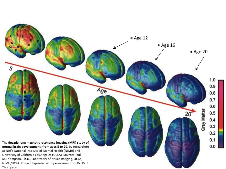
Script error: No such module "Navbox". Script error: No such module "Navbox". Template:Authority control